Itoh Yuta, Hayakawa Kimihide, Mori Tomohiro, Agata Nobuhide, Inoue-Miyazu Masumi, Murakami Taro, Sokabe Masahiro, Kawakami Keisuke
Physical and Occupational Therapy Program, Nagoya University Graduate School of Medicine, Nagoya, Japan Faculty of Rehabilitation Science, Nagoya Gakuin University, Seto, Japan.
Mechanobiology Laboratory, Nagoya University Graduate School of Medicine, Nagoya, Japan.
Physiol Rep. 2014 Nov 3;2(11). doi: 10.14814/phy2.12185. Print 2014 Nov 1.
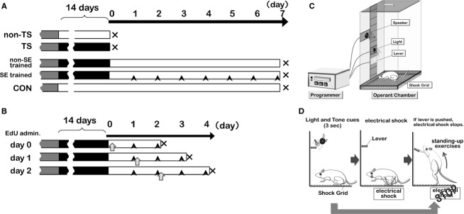
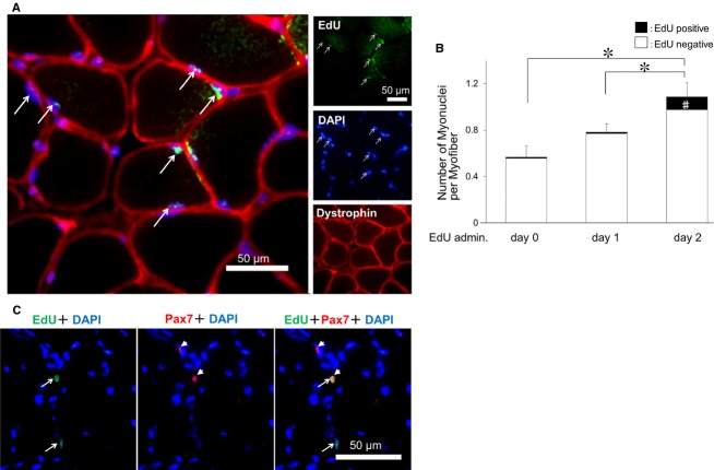
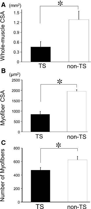
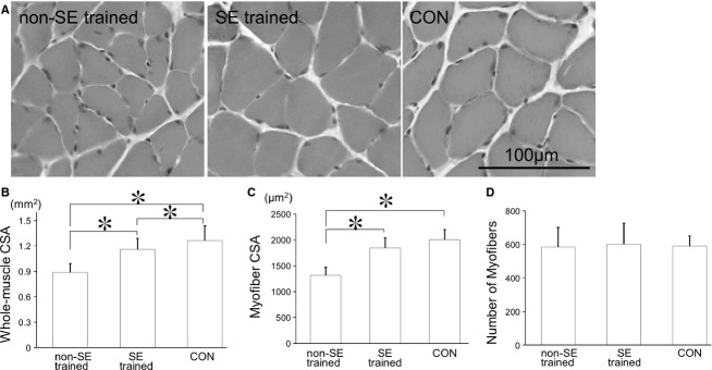
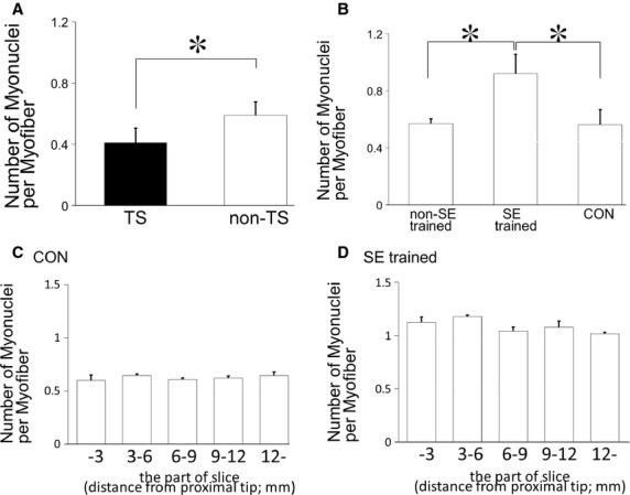
Determining the cellular and molecular recovery processes in inactivity - or unloading -induced atrophied muscles should improve rehabilitation strategies. We assessed the effects of stand-up exercise (SE) training on the recovery of atrophied skeletal muscles in male mice. Mice were trained to stand up and press an elevated lever in response to a light-tone cue preceding an electric foot shock and then subjected to tail suspension (TS) for 2 weeks to induce disuse atrophy in hind limb muscles. After release from TS, mice were divided into SE-trained (SE cues: 25 times per set, two sets per day) and non-SE-trained groups. Seven days after the training, average myofiber cross-sectional area (CSA) of the soleus muscle was significantly greater in the SE-trained group than in the non-SE-trained group (1843 ± 194 μm(2) vs. 1315 ± 153 μm(2)). Mean soleus muscle CSA in the SE trained group was not different from that in the CON group subjected to neither TS nor SE training (2005 ± 196 μm(2)), indicating that SE training caused nearly complete recovery from muscle atrophy. The number of myonuclei per myofiber was increased by ~60% in the SE-trained group compared with the non-SE-trained and CON groups (0.92 ± 0.03 vs. 0.57 ± 0.03 and 0.56 ± 0.11, respectively). The number of proliferating myonuclei, identified by 5-ethynyl-2'-deoxyuridine staining, increased within the first few days of SE training. Thus, it is highly likely that myogenic satellite cells proliferated rapidly in atrophied muscles in response to SE training and fused with existing myofibers to reestablish muscle mass.
确定不活动(或卸载)诱导的萎缩肌肉中的细胞和分子恢复过程,应能改善康复策略。我们评估了站立运动(SE)训练对雄性小鼠萎缩骨骼肌恢复的影响。训练小鼠在电足电击前的轻音提示下站立并按压升高的杠杆,然后使其尾部悬吊(TS)2周,以诱导后肢肌肉废用性萎缩。从TS解除后,将小鼠分为SE训练组(SE提示:每组25次,每天两组)和非SE训练组。训练7天后,SE训练组比目鱼肌的平均肌纤维横截面积(CSA)显著大于非SE训练组(1843±194μm²对1315±153μm²)。SE训练组的比目鱼肌平均CSA与既未接受TS也未接受SE训练的CON组(2005±196μm²)无差异,表明SE训练使肌肉萎缩几乎完全恢复。与非SE训练组和CON组相比,SE训练组每根肌纤维的肌核数量增加了约60%(分别为0.92±0.03对0.57±0.03和0.56±0.11)。通过5-乙炔基-2'-脱氧尿苷染色鉴定的增殖肌核数量在SE训练的头几天内增加。因此,很有可能成肌卫星细胞在萎缩肌肉中因SE训练而迅速增殖,并与现有的肌纤维融合以重建肌肉质量。