Janssen Rob, Zuidwijk Marian J, Kuster Diederik W D, Muller Alice, Simonides Warner S
Department of Physiology, VU University Medical Center, Institute for Cardiovascular Research , Amsterdam , Netherlands.
Front Endocrinol (Lausanne). 2014 Oct 20;5:171. doi: 10.3389/fendo.2014.00171. eCollection 2014.
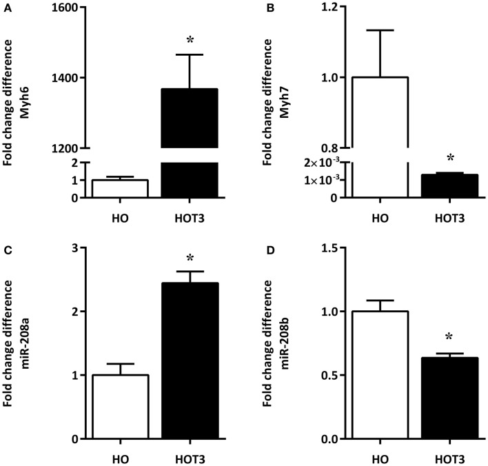
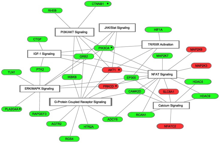
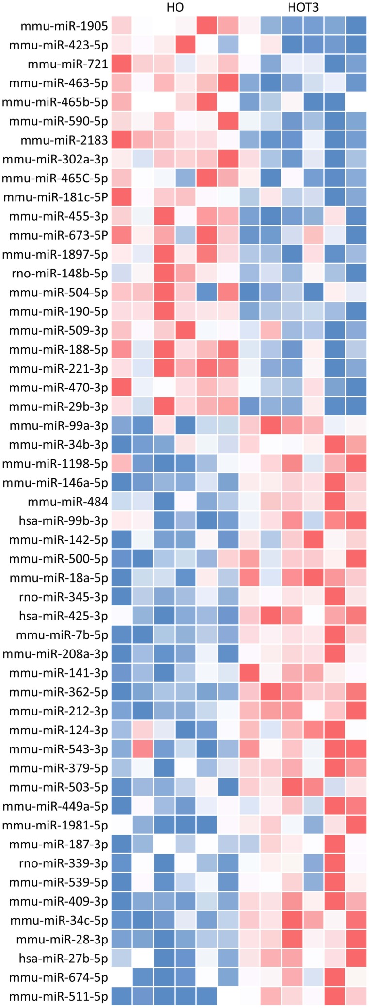
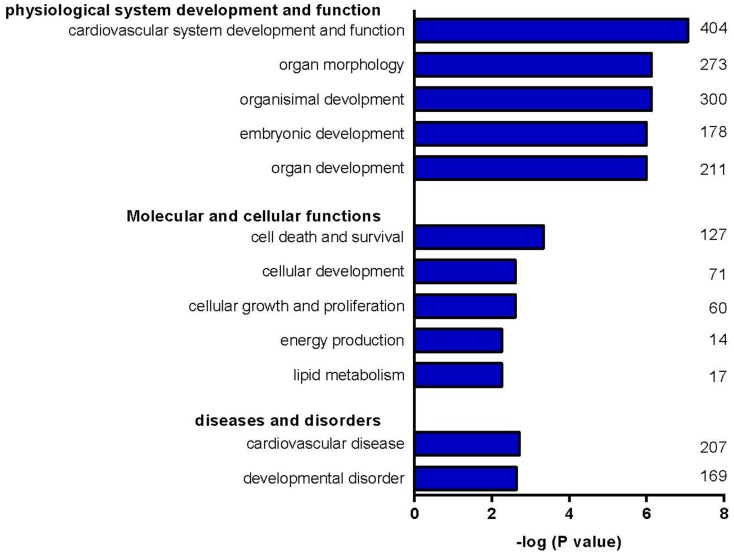
Cardiomyocyte size in the healthy heart is in part determined by the level of circulating thyroid hormone (TH). Higher levels of TH induce ventricular hypertrophy, primarily in response to an increase in hemodynamic load. Normal cardiac function is maintained in this form of hypertrophy, whereas progressive contractile dysfunction is a hallmark of pathological hypertrophy. MicroRNAs (miRNAs) are important modulators of signal-transduction pathways driving adverse remodeling. Because little is known about the involvement of miRNAs in cardiac TH action and hypertrophy, we examined the miRNA expression profile of the hypertrophied left ventricle (LV) using a mouse model of TH-induced cardiac hypertrophy. C57Bl/6J mice were rendered hypothyroid by treatment with propylthiouracil and were subsequently treated for 3 days with TH (T3) or saline. T3 treatment increased LV weight by 38% (p < 0.05). RNA was isolated from the LV and expression of 641 mouse miRNAs was determined using Taqman Megaplex arrays. Data were analyzed using RQ-manager and DataAssist. A total of 52 T3-regulated miRNAs showing a >2-fold change (p < 0.05) were included in Ingenuity Pathway Analysis to predict target mRNAs involved in cardiac hypertrophy. The analysis was further restricted to proteins that have been validated as key factors in hypertrophic signal transduction in mouse models of ventricular remodeling. A total of 27 mRNAs were identified as bona fide targets. The predicted regulation of 19% of these targets indicates enhancement of physiological hypertrophy, while 56% indicates suppression of pathological remodeling. Our data suggest that cardiac TH action includes a novel level of regulation in which a unique set of TH-dependent miRNAs primarily suppresses pathological hypertrophic signaling. This may be relevant for our understanding of the progression of adverse remodeling, since cardiac TH levels are known to decrease substantially in various forms of pathological hypertrophy.
健康心脏中心肌细胞的大小部分由循环甲状腺激素(TH)水平决定。较高水平的TH会诱发心室肥大,主要是对血流动力学负荷增加的反应。这种肥大形式可维持正常心脏功能,而进行性收缩功能障碍是病理性肥大的标志。微小RNA(miRNA)是驱动不良重塑的信号转导通路的重要调节因子。由于对miRNA在心脏TH作用和肥大中的参与情况了解甚少,我们使用TH诱导的心脏肥大小鼠模型研究了肥厚左心室(LV)的miRNA表达谱。通过丙硫氧嘧啶治疗使C57Bl/6J小鼠甲状腺功能减退,随后用TH(T3)或生理盐水治疗3天。T3治疗使LV重量增加了38%(p<0.05)。从LV中分离RNA,并使用Taqman Megaplex阵列测定641种小鼠miRNA的表达。使用RQ-manager和DataAssist分析数据。共有52种受T3调节的miRNA显示出>2倍的变化(p<0.05),被纳入 Ingenuity Pathway Analysis以预测参与心脏肥大的靶mRNA。分析进一步局限于已被证实为心室重塑小鼠模型中肥厚信号转导关键因子的蛋白质。总共鉴定出27种mRNA为真正的靶标。这些靶标中19%的预测调节表明生理肥大增强,而56%表明病理性重塑受到抑制。我们的数据表明,心脏TH作用包括一个新的调节水平,其中一组独特的TH依赖性miRNA主要抑制病理性肥厚信号。这可能与我们对不良重塑进展的理解相关,因为已知在各种形式的病理性肥大中,心脏TH水平会大幅下降。