School of Medicine and Medical Science, University College Dublin, Dublin, 4 Ireland.
Conway Institute, University College Dublin, Dublin, 4 Ireland.
Cancer Cell Int. 2014 Oct 30;14(1):108. doi: 10.1186/s12935-014-0108-6. eCollection 2014.
The cancer microenvironment has a strong impact on the growth and dynamics of cancer cells. Conventional 2D culture systems, however, do not reflect in vivo conditions, impeding detailed studies of cancer cell dynamics. This work aims to establish a method to reveal the interaction of cancer and normal epithelial cells using 3D time-lapse.
GFP-labelled breast cancer cells, MDA-MB-231, were co-cultured with mCherry-labelled non-cancerous epithelial cells, MDCK, in a gel matrix. In the 3D culture, the epithelial cells establish a spherical morphology (epithelial sphere) thus providing cancer cells with accessibility to the basal surface of epithelia, similar to the in vivo condition. Cell movement was monitored using time-lapse analyses. Ultrastructural, immunocytochemical and protein expression analyses were also performed following the time-lapse study.
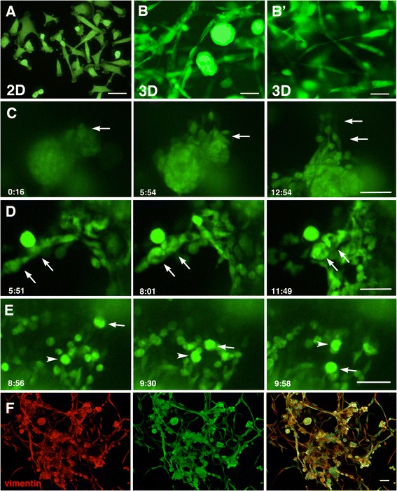
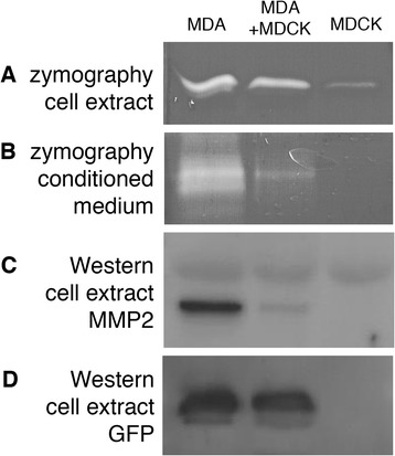
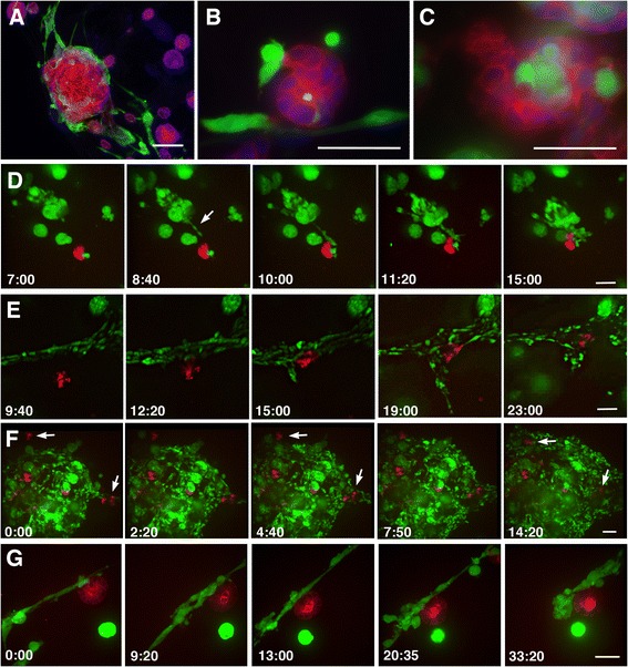
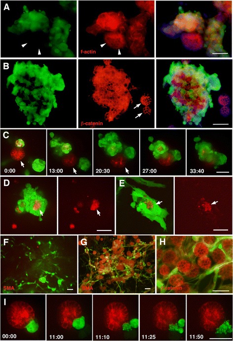
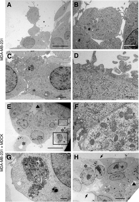
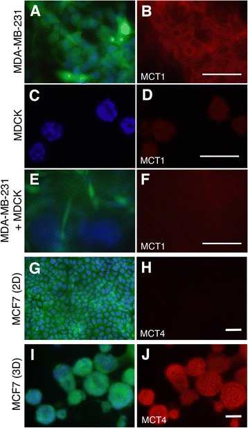
In contrast to the 2D culture system, whereby most MDA-MB-231 cells exhibit spindle-shaped morphology as single cells, in the 3D culture the MDA-MB-231 cells were found to be single cells or else formed aggregates, both of which were motile. The single MDA-MB-231 cells exhibited both round and spindle shapes, with dynamic changes from one shape to the other, visible within a matter of hours. When co-cultured with epithelial cells, the MDA-MB-231 cells displayed a strong attraction to the epithelial spheres, and proceeded to surround and engulf the epithelial cell mass. The surrounded epithelial cells were eventually destroyed, becoming debris, and were taken into the MDA-MB-231 cells. However, when there was a relatively large population of normal epithelial cells, the MDA-MB-231 cells did not engulf the epithelial spheres effectively, despite repeated contacts. MDA-MB-231 cells co-cultured with a large number of normal epithelial cells showed reduced expression of monocarboxylate transporter-1, suggesting a change in the cell metabolism. A decreased level of gelatin-digesting ability as well as reduced production of matrix metaroproteinase-2 was also observed.
This culture method is a powerful technique to investigate cancer cell dynamics and cellular changes in response to the microenvironment. The method can be useful for various aspects such as; different combinations of cancer and non-cancer cell types, addressing the organ-specific affinity of cancer cells to host cells, and monitoring the cellular response to anti-cancer drugs.
癌症微环境对癌细胞的生长和动态有很强的影响。然而,传统的 2D 培养系统不能反映体内条件,阻碍了对癌细胞动态的详细研究。本工作旨在建立一种使用 3D 延时拍摄揭示癌症和正常上皮细胞相互作用的方法。
用 GFP 标记的乳腺癌细胞 MDA-MB-231 与 mCherry 标记的非癌上皮细胞 MDCK 共培养在凝胶基质中。在 3D 培养中,上皮细胞形成球形形态(上皮球体),从而使癌细胞能够接近上皮的基底表面,类似于体内条件。使用延时分析监测细胞运动。延时研究后还进行了超微结构、免疫细胞化学和蛋白表达分析。
与 2D 培养系统相比,大多数 MDA-MB-231 细胞在 2D 培养系统中呈纺锤形形态作为单细胞,而在 3D 培养系统中,MDA-MB-231 细胞发现是单细胞或聚集在一起,这两种状态都是可移动的。单个 MDA-MB-231 细胞呈现圆形和纺锤形两种形态,在数小时内可见从一种形态到另一种形态的动态变化。当与上皮细胞共培养时,MDA-MB-231 细胞对上皮球体有强烈的吸引力,并开始包围和吞噬上皮细胞团。被包围的上皮细胞最终被破坏,成为碎片,并被 MDA-MB-231 细胞吸收。然而,当有大量正常上皮细胞时,尽管反复接触,MDA-MB-231 细胞也不能有效地吞噬上皮球体。与大量正常上皮细胞共培养的 MDA-MB-231 细胞表达的单羧酸转运蛋白-1 减少,表明细胞代谢发生变化。还观察到明胶消化能力降低和基质金属蛋白酶-2 产生减少。
这种培养方法是研究癌细胞动态和细胞对微环境响应变化的有力技术。该方法可用于各种方面,例如不同类型的癌症和非癌细胞的组合,解决癌细胞对宿主细胞的器官特异性亲和力,以及监测细胞对抗癌药物的反应。