Usta Ufuk, Tastekin Ebru, Isler Erhan, Kutlu Ali K, Oz Puyan Fulya
Department of Pathology, Medical Faculty, Trakya University, Edirne, Turkey. Tel. +90 (284) 2357641/2351528. Fax. +90 (284) 2352730. E-mail.
Saudi Med J. 2014 Nov;35(11):1331-8.
To collect data on all detectable histologic and immune alterations from the kidneys of 55 autopsy cases.
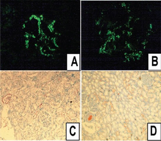
This prospective study was carried out at the Department of Pathology, Medical Faculty, Trakya University, Edirne, Turkey. Fifty-five cases were subjected to the study among 248 autopsies that were performed in 2011 and 2012. All kidney samples were evaluated under a light microscope and fresh tissue samples were used for immunofluorescence microscopy. Immunohistochemically kappa (κ) and lambda (λ) antibodies were applied to the tissue sections. The glomerular, tubulo-interstitial, and vascular alterations, as well as immune depositions were noted.
The microscopic morphology was close to normal histology in only 23 cases, and 23 cases had glomerular alterations. Nineteen cases had at least one immune deposition. There was immunoglobulin A deposition in 13 cases, and 9 cases showed positivity for both κ and λ immunohistochemically, and there was no clonal positivity.
The most striking outcome of our study is the high rate of immune depositions. There was also a significant number of glomerular and non-glomerular renal alterations.
收集55例尸检病例肾脏所有可检测到的组织学和免疫改变的数据。
这项前瞻性研究在土耳其埃迪尔内特拉凯亚大学医学院病理科进行。在2011年和2012年进行的248例尸检中,有55例纳入研究。所有肾脏样本均在光学显微镜下评估,新鲜组织样本用于免疫荧光显微镜检查。免疫组织化学方法将κ和λ抗体应用于组织切片。记录肾小球、肾小管间质和血管的改变以及免疫沉积物。
仅23例微观形态接近正常组织学,23例有肾小球改变。19例至少有一处免疫沉积物。13例有免疫球蛋白A沉积,9例κ和λ免疫组织化学均呈阳性,且无克隆性阳性。
我们研究最显著的结果是免疫沉积物的高发生率。也有大量的肾小球和非肾小球性肾脏改变。