Itoh Akihiro, Uchiyama Atsushi, Taniguchi Shunichiro, Sagara Junji
Department of Biomedical Laboratory Sciences, Health Sciences, Shinshu University, Matsumoto, Japan.
Department of Molecular Oncology, Medical Sciences, Shinshu University Graduate School of Medicine, Matsumoto, Japan.
PLoS One. 2014 Nov 18;9(11):e113289. doi: 10.1371/journal.pone.0113289. eCollection 2014.
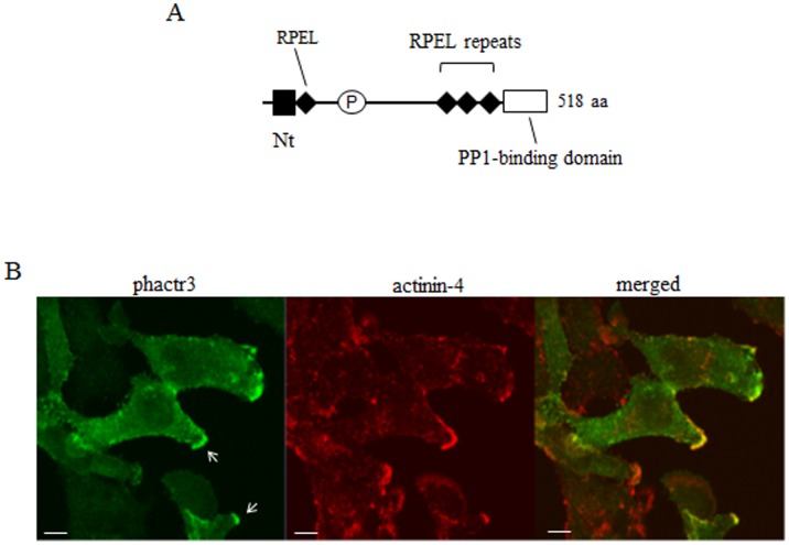
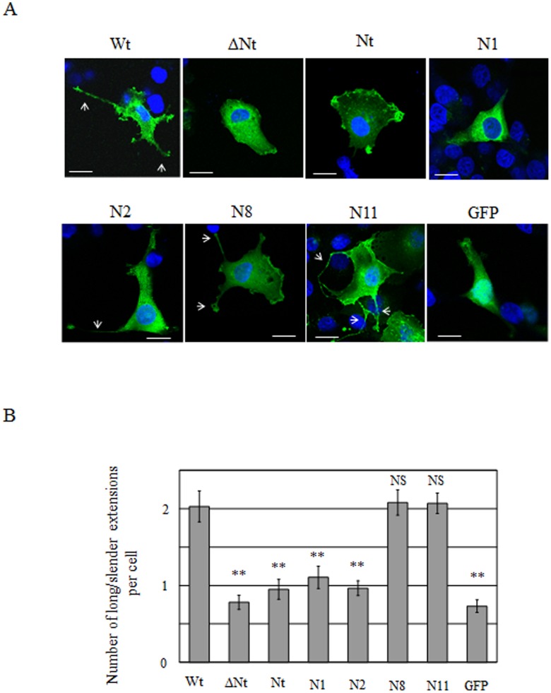
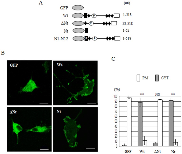
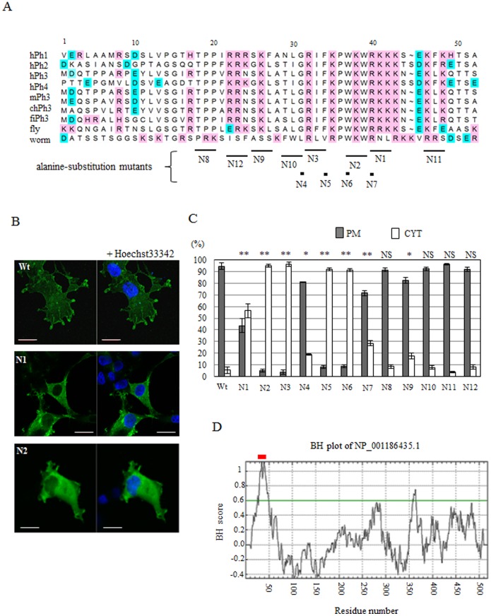
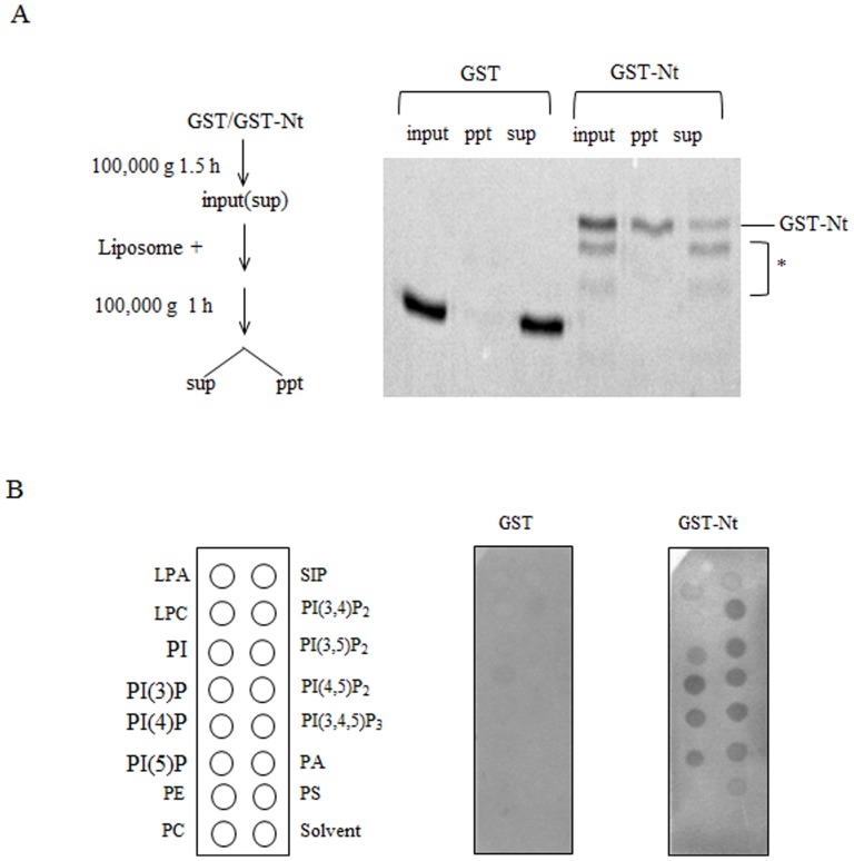
Proteins that belong to the protein phosphatase 1 and actin regulator (phactr) family are involved in cell motility and morphogenesis. However, the mechanisms that regulate the actin cytoskeleton are poorly understood. We have previously shown that phactr3, also known as scapinin, localizes to the plasma membrane, including lamellipodia and membrane ruffles. In the present study, experiments using deletion and point mutants showed that the basic and hydrophobic residues in the N-terminus play crucial roles in the localization to the plasma membrane. A BH analysis (http://helixweb.nih.gov/bhsearch) is a program developed to identify membrane-binding domains that comprise basic and hydrophobic residues in membrane proteins. We applied this program to phactr3. The results of the BH plot analysis agreed with the experimentally determined region that is responsible for the localization of phactr3 to the plasma membrane. In vitro experiments showed that the N-terminal itself binds to liposomes and acidic phospholipids. In addition, we showed that the interaction with the plasma membrane via the N-terminal membrane-binding sequence is required for phactr3-induced morphological changes in Cos7 cells. The membrane-binding sequence in the N-terminus is highly conserved in all members of the phactr family. Our findings may provide a molecular basis for understanding the mechanisms that allow phactr proteins to regulate cell morphogenesis.
属于蛋白磷酸酶1和肌动蛋白调节剂(phactr)家族的蛋白质参与细胞运动和形态发生。然而,调节肌动蛋白细胞骨架的机制仍知之甚少。我们之前已经表明,phactr3,也称为scapinin,定位于质膜,包括片状伪足和膜皱褶。在本研究中,使用缺失和点突变体的实验表明,N端的碱性和疏水残基在质膜定位中起关键作用。BH分析(http://helixweb.nih.gov/bhsearch)是一个开发用于识别膜蛋白中包含碱性和疏水残基的膜结合结构域的程序。我们将此程序应用于phactr3。BH图分析的结果与实验确定的负责phactr3定位于质膜的区域一致。体外实验表明,N端本身与脂质体和酸性磷脂结合。此外,我们表明,通过N端膜结合序列与质膜的相互作用是phactr3诱导Cos7细胞形态变化所必需的。N端的膜结合序列在phactr家族的所有成员中高度保守。我们的发现可能为理解phactr蛋白调节细胞形态发生的机制提供分子基础。