Genevini Paola, Papiani Giulia, Ruggiano Annamaria, Cantoni Lavinia, Navone Francesca, Borgese Nica
Institute of Neuroscience, Consiglio Nazionale delle Ricerche, and Department of Medical Biotechnology and Translational Medicine (BIOMETRA), Università degli Studi di Milano, Milano, Italy.
Department of Molecular Biochemistry and Pharmacology, Istituto di Ricerche Farmacologiche "Mario Negri", Milan, Italy.
PLoS One. 2014 Nov 19;9(11):e113416. doi: 10.1371/journal.pone.0113416. eCollection 2014.
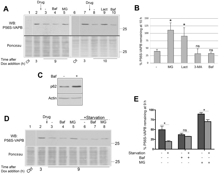
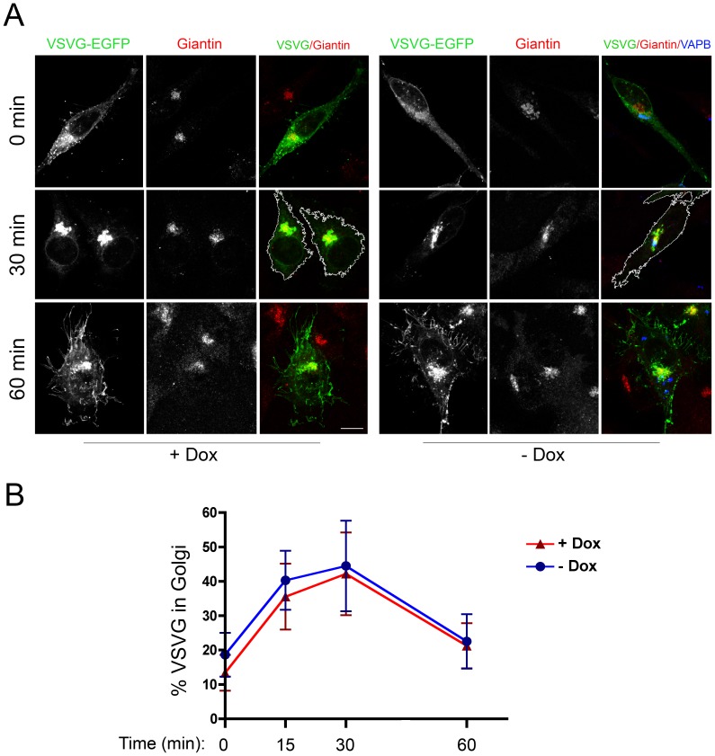
VAPB is a ubiquitously expressed, ER-resident adaptor protein involved in interorganellar lipid exchange, membrane contact site formation, and membrane trafficking. Its mutant form, P56S-VAPB, which has been linked to a dominantly inherited form of Amyotrophic Lateral Sclerosis (ALS8), generates intracellular inclusions consisting in restructured ER domains whose role in ALS pathogenesis has not been elucidated. P56S-VAPB is less stable than the wild-type protein and, at variance with most pathological aggregates, its inclusions are cleared by the proteasome. Based on studies with cultured cells overexpressing the mutant protein, it has been suggested that VAPB inclusions may exert a pathogenic effect either by sequestering the wild-type protein and other interactors (loss-of-function by a dominant negative effect) or by a more general proteotoxic action (gain-of-function). To investigate P56S-VAPB degradation and the effect of the inclusions on proteostasis and on ER-to-plasma membrane protein transport in a more physiological setting, we used stable HeLa and NSC34 Tet-Off cell lines inducibly expressing moderate levels of P56S-VAPB. Under basal conditions, P56S-VAPB degradation was mediated exclusively by the proteasome in both cell lines, however, it could be targeted also by starvation-stimulated autophagy. To assess possible proteasome impairment, the HeLa cell line was transiently transfected with the ERAD (ER Associated Degradation) substrate CD3δ, while autophagic flow was investigated in cells either starved or treated with an autophagy-stimulating drug. Secretory pathway functionality was evaluated by analyzing the transport of transfected Vesicular Stomatitis Virus Glycoprotein (VSVG). P56S-VAPB expression had no effect either on the degradation of CD3δ or on the levels of autophagic markers, or on the rate of transport of VSVG to the cell surface. We conclude that P56S-VAPB inclusions expressed at moderate levels do not interfere with protein degradation pathways or protein transport, suggesting that the dominant inheritance of the mutant gene may be due mainly to haploinsufficiency.
VAPB是一种广泛表达的内质网驻留衔接蛋白,参与细胞器间脂质交换、膜接触位点形成和膜运输。其突变形式P56S-VAPB与一种显性遗传形式的肌萎缩侧索硬化症(ALS8)相关,会产生由重组内质网结构域组成的细胞内包涵体,其在ALS发病机制中的作用尚未阐明。P56S-VAPB比野生型蛋白稳定性差,与大多数病理性聚集体不同,其包涵体可被蛋白酶体清除。基于对过表达突变蛋白的培养细胞的研究,有人提出VAPB包涵体可能通过隔离野生型蛋白和其他相互作用分子(通过显性负效应导致功能丧失)或通过更普遍的蛋白毒性作用(功能获得)发挥致病作用。为了在更接近生理的环境中研究P56S-VAPB的降解以及包涵体对蛋白质稳态和内质网到质膜蛋白运输的影响,我们使用了稳定的HeLa和NSC34 Tet-Off细胞系,可诱导表达中等水平的P56S-VAPB。在基础条件下,P56S-VAPB的降解在两种细胞系中均仅由蛋白酶体介导,然而,饥饿刺激的自噬也可将其作为靶点。为了评估可能的蛋白酶体损伤,HeLa细胞系用内质网相关降解(ERAD)底物CD3δ进行瞬时转染,同时在饥饿或用自噬刺激药物处理的细胞中研究自噬流。通过分析转染的水疱性口炎病毒糖蛋白(VSVG)的运输来评估分泌途径功能。P56S-VAPB的表达对CD3δ的降解、自噬标志物水平或VSVG向细胞表面的运输速率均无影响。我们得出结论,中等水平表达的P56S-VAPB包涵体不会干扰蛋白质降解途径或蛋白质运输,这表明突变基因的显性遗传可能主要归因于单倍体不足。