Plantinga Birgit R, Temel Yasin, Roebroeck Alard, Uludağ Kâmil, Ivanov Dimo, Kuijf Mark L, Ter Haar Romenij Bart M
Biomedical Image Analysis, Eindhoven University of Technology Eindhoven, Netherlands ; Department of Neuroscience, Maastricht University Maastricht, Netherlands.
Department of Neuroscience, Maastricht University Maastricht, Netherlands ; Department of Neurology, Maastricht University Medical Center Maastricht, Netherlands.
Front Hum Neurosci. 2014 Nov 5;8:876. doi: 10.3389/fnhum.2014.00876. eCollection 2014.
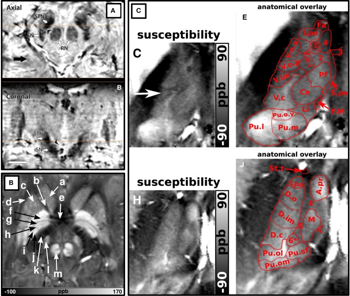
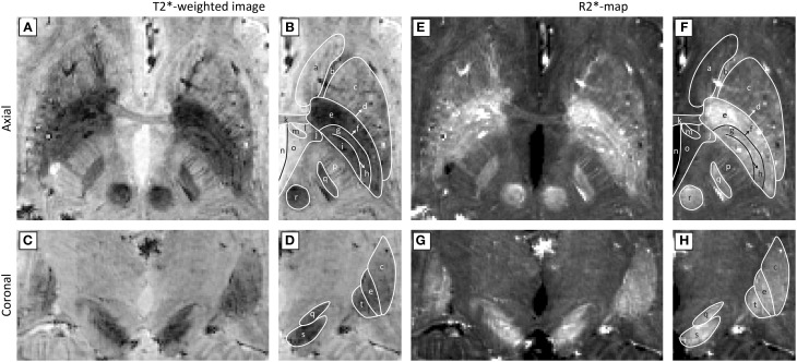
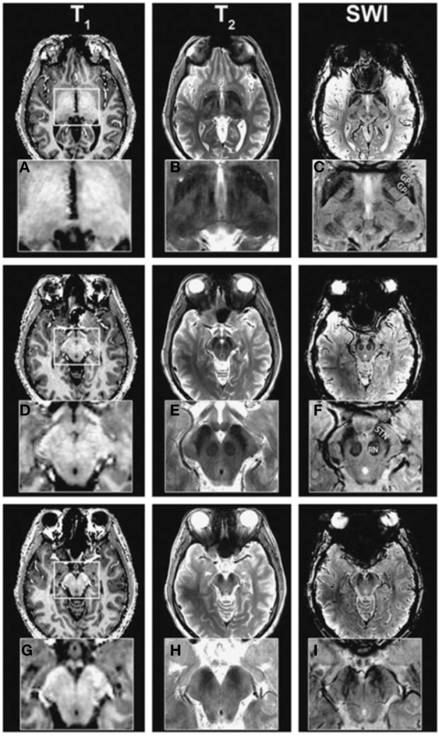
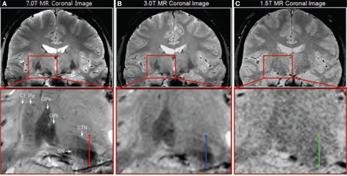
Deep brain stimulation is a treatment for Parkinson's disease and other related disorders, involving the surgical placement of electrodes in the deeply situated basal ganglia or thalamic structures. Good clinical outcome requires accurate targeting. However, due to limited visibility of the target structures on routine clinical MR images, direct targeting of structures can be challenging. Non-clinical MR scanners with ultra-high magnetic field (7T or higher) have the potential to improve the quality of these images. This technology report provides an overview of the current possibilities of visualizing deep brain stimulation targets and their related structures with the aid of ultra-high field MRI. Reviewed studies showed improved resolution, contrast- and signal-to-noise ratios at ultra-high field. Sequences sensitive to magnetic susceptibility such as T2(*) and susceptibility weighted imaging and their maps in general showed the best visualization of target structures, including a separation between the subthalamic nucleus and the substantia nigra, the lamina pallidi medialis and lamina pallidi incompleta within the globus pallidus and substructures of the thalamus, including the ventral intermediate nucleus (Vim). This shows that the visibility, identification, and even subdivision of the small deep brain stimulation targets benefit from increased field strength. Although ultra-high field MR imaging is associated with increased risk of geometrical distortions, it has been shown that these distortions can be avoided or corrected to the extent where the effects are limited. The availability of ultra-high field MR scanners for humans seems to provide opportunities for a more accurate targeting for deep brain stimulation in patients with Parkinson's disease and related disorders.
深部脑刺激是治疗帕金森病及其他相关疾病的一种方法,涉及将电极手术植入深部的基底神经节或丘脑结构。良好的临床效果需要精确的靶点定位。然而,由于常规临床磁共振成像(MR)图像上目标结构的可视性有限,直接对结构进行靶向定位可能具有挑战性。具有超高磁场(7T或更高)的非临床MR扫描仪有潜力提高这些图像的质量。本技术报告概述了借助超高场MRI可视化深部脑刺激靶点及其相关结构的当前可能性。综述研究表明,在超高场下分辨率、对比度和信噪比均有所提高。对磁敏感性敏感的序列,如T2(*)和磁敏感加权成像及其图谱,总体上对目标结构的可视化效果最佳,包括丘脑底核与黑质之间的分离、苍白球内侧板和苍白球不完全板在苍白球内的显示,以及丘脑的亚结构,包括腹中间核(Vim)。这表明,增加场强有利于提高深部脑刺激小靶点的可视性、识别性,甚至可进行细分。尽管超高场MR成像会增加几何畸变的风险,但已表明这些畸变可以避免或校正到影响有限的程度。可供人类使用的超高场MR扫描仪似乎为帕金森病及相关疾病患者进行更精确的深部脑刺激靶点定位提供了机会。