Department of Neurology and Neurological Sciences, Stanford University School of Medicine, Stanford, CA, USA.
School of Medicine, University of Massachusetts, Worcester, MA, USA.
J Parkinsons Dis. 2020;10(2):591-604. doi: 10.3233/JPD-191890.
In postmortem analysis of late stage Parkinson's disease (PD) neuronal loss in the substantial nigra (SN) correlates with the antemortem severity of bradykinesia and rigidity, but not tremor.
To investigate the relationship between midbrain nuclei volume as an in vivo biomarker for surviving neurons in mild-to-moderate patients using 7.0 Tesla MRI.
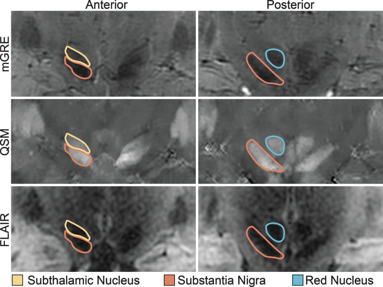
We performed ultra-high resolution quantitative susceptibility mapping (QSM) on the midbrain in 32 PD participants with less than 10 years duration and 8 healthy controls. Following blinded manual segmentation, the individual volumes of the SN, subthalamic nucleus, and red nucleus were measured. We then determined the associations between the midbrain nuclei and clinical metrics (age, disease duration, MDS-UPDRS motor score, and subscores for bradykinesia/rigidity, tremor, and postural instability/gait difficulty).
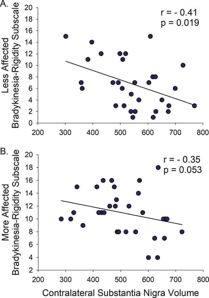
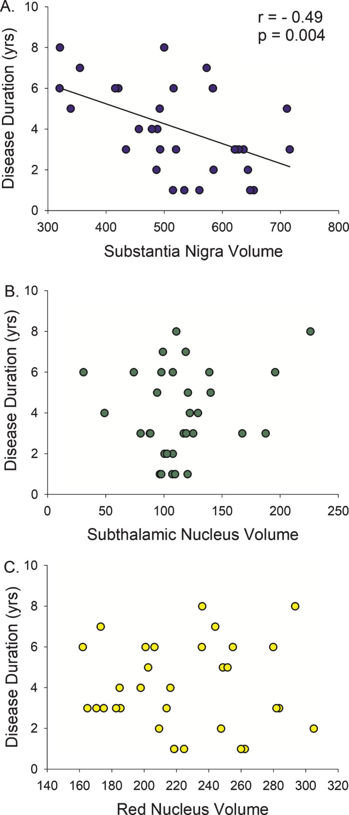
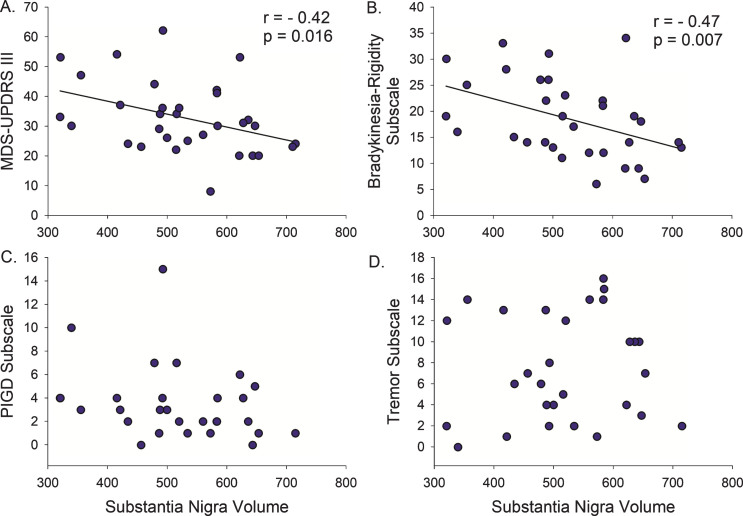
We found that smaller SN correlated with longer disease duration (r = -0.49, p = 0.004), more severe MDS-UPDRS motor score (r = -0.42, p = 0.016), and more severe bradykinesia-rigidity subscore (r = -0.47, p = 0.007), but not tremor or postural instability/gait difficulty subscores. In a hemi-body analysis, bradykinesia-rigidity severity only correlated with SN contralateral to the less-affected hemi-body, and not contralateral to the more-affected hemi-body, possibly reflecting the greatest change in dopamine neuron loss early in disease. Multivariate generalized estimating equation model confirmed that bradykinesia-rigidity severity, age, and disease duration, but not tremor severity, predicted SN volume.
In mild-to-moderate PD, SN volume relates to motor manifestations in a motor domain-specific and laterality-dependent manner. Non-invasive in vivo 7.0 Tesla QSM may serve as a biomarker in longitudinal studies of SN atrophy and in studies of people at risk for developing PD.
在晚期帕金森病(PD)的死后分析中,黑质(SN)中的神经元丧失与生前运动迟缓的严重程度相关,但与震颤无关。
使用 7.0T MRI 研究中脑核体积作为轻度至中度患者存活神经元的体内生物标志物的关系。
我们对 32 名病程不足 10 年的 PD 患者和 8 名健康对照者进行了中脑超高分辨率定量磁化率映射(QSM)。在进行盲法手动分割后,测量 SN、丘脑下核和红核的个体体积。然后,我们确定了中脑核与临床指标(年龄、病程、MDS-UPDRS 运动评分以及运动迟缓/僵硬、震颤和姿势不稳/步态困难的亚评分)之间的关系。
我们发现,SN 越小与病程越长(r=-0.49,p=0.004)、MDS-UPDRS 运动评分越高(r=-0.42,p=0.016)、运动迟缓/僵硬亚评分越高(r=-0.47,p=0.007)相关,但与震颤或姿势不稳/步态困难亚评分无关。在半身分析中,运动迟缓/僵硬严重程度仅与受影响较小的半身对侧的 SN 相关,而与受影响较大的半身的 SN 无关,这可能反映了疾病早期多巴胺神经元丧失的最大变化。多变量广义估计方程模型证实,运动迟缓/僵硬严重程度、年龄和病程,而不是震颤严重程度,预测了 SN 体积。
在轻度至中度 PD 中,SN 体积与运动表现相关,具有运动域特异性和侧别依赖性。7.0T QSM 可能成为 SN 萎缩的纵向研究以及 PD 高危人群研究中的生物标志物。