Lockard R E, Alzner-Deweerd B, Heckman J E, MacGee J, Tabor M W, RajBhandary U L
Nucleic Acids Res. 1978 Jan;5(1):37-56. doi: 10.1093/nar/5.1.37.
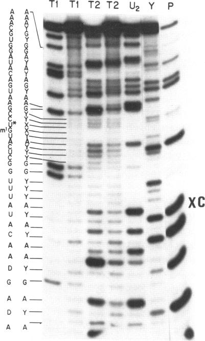
Sequence analysis of 5'-[32P] labeled tRNA and eukaryotic mRNA using an adaptation of a method recently described by Donis-Keller, Maxam and Gilbert for mapping guanines, adenines and pyrimidines from the 5'-end of an RNA is described. In addition, a technique utilizing two-dimensional polyacrylamide gel electrophoresis for identification of pyrimidines within a sequence is described. 5'-[32P] Labeled rabbit beta-globin mRNA and N. crassa mitochondrial initiator tRNA were partially digested with T1- RNase for cleavage at G residues, with U2-RNase for cleavage at A residues, with an extracellular RNase from B. cereus for cleavage at pyrimidine residues and with T2-RNase or with alkali for cleavage at all four residues. The 5'-[32P] labeled partial digestion products were separated according to their size, by electrophoresis in adjacent lanes of a polyacrylamide slab gel and the location of G's, A's and of pyrimidines extending 60-80 nucleotides from the 5'-end of the RNA determined. Two-dimensional polyacrylamide gel electrophoresis was used to separate the 5'-[32P] labeled fragments present in partial alkali digests of a 5'-[32P] labeled mRNA. The mobility shifts corresponding to the difference of a C residue were distinct from those corresponding to a U residue and this formed the basis of a method for distinguishing between the pyrimidines.
本文描述了使用一种改编自多尼斯 - 凯勒、马克萨姆和吉尔伯特最近描述的用于从RNA 5'端定位鸟嘌呤、腺嘌呤和嘧啶的方法,对5'-[³²P]标记的tRNA和真核mRNA进行序列分析。此外,还描述了一种利用二维聚丙烯酰胺凝胶电泳来鉴定序列中嘧啶的技术。5'-[³²P]标记的兔β-珠蛋白mRNA和粗糙脉孢菌线粒体起始tRNA分别用T1核糖核酸酶在G残基处切割、用U2核糖核酸酶在A残基处切割、用蜡状芽孢杆菌的细胞外核糖核酸酶在嘧啶残基处切割,以及用T2核糖核酸酶或碱在所有四个残基处切割进行部分消化。5'-[³²P]标记的部分消化产物根据其大小,在聚丙烯酰胺平板凝胶的相邻泳道中通过电泳进行分离,并确定从RNA 5'端延伸60 - 80个核苷酸的G、A和嘧啶的位置。二维聚丙烯酰胺凝胶电泳用于分离5'-[³²P]标记的mRNA部分碱消化中存在的5'-[³²P]标记片段。对应于C残基差异的迁移率变化与对应于U残基的迁移率变化不同,这构成了区分嘧啶的一种方法的基础。