Kim Min Hee, Hwang Ho Sik, Park Kyoung Jin, Hwang Je Hyung, Joo Choun Ki
Department of Ophthalmology and Visual Science, Seoul St. Mary's Hospital, The Catholic University of Korea College of Medicine, Seoul, Korea.
Department of Ophthalmology, Chuncheon Sacred Heart Hospital, Hallym University College of Medicine, Chuncheon, Korea.
Korean J Ophthalmol. 2014 Dec;28(6):486-92. doi: 10.3341/kjo.2014.28.6.486. Epub 2014 Nov 19.
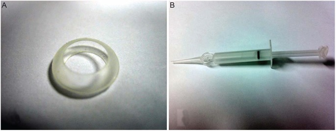
In this study, we examined the stability of the lens-angle supporter (LAS) for accommodation restoration by comparing intraocular lens (IOL) location, after-cataract and ciliary body damage after cataract surgery in rabbits.
Eight rabbits were divided into experimental and control groups of four rabbits each. Phacoemulsification and irrigation and aspiration were performed in all rabbits. This was followed by an LAS and IOL insertion in the four experimental rabbits. In the four control rabbits, only an IOL insertion was performed. Six months after the surgery, the location of the IOL, the conditions of the lens capsule and ciliary body were evaluated using a slitl-amp examination and Miyake-Apple view.
For the experimental group, the ultrasound biomicroscope results showed normal LAS and IOL positioning in all four cases. According to the slitlamp examination and Miyake-Apple view, the IOL was positioned at the center, with less after-cataract and damage to the ciliary body. For the control group, ultrasound biomicroscope results indicated a higher IOL position than normal, as well as a single case of IOL decentering. According to the slit-lamp examination and Miyake-Apple view, the IOL was decentered with more severe after-cataract and ciliary body damage.
The LAS has the potential to maintain a stable IOL position while producing less after-cataract when used in lens-angle reconstruction for correction of presbyopia. Moreover, LAS implantation incurs less damage to the ciliary body.
在本研究中,我们通过比较兔白内障手术后人工晶状体(IOL)的位置、后发性白内障及睫状体损伤情况,研究晶状体角支撑体(LAS)用于恢复调节功能的稳定性。
将8只兔分为实验组和对照组,每组4只。所有兔均行超声乳化及灌吸术。之后,4只实验兔植入LAS和IOL,4只对照兔仅植入IOL。术后6个月,使用裂隙灯检查和Miyake-Apple视图评估IOL的位置、晶状体囊膜及睫状体的情况。
实验组中,超声生物显微镜检查结果显示所有4例LAS和IOL定位均正常。根据裂隙灯检查和Miyake-Apple视图,IOL位于中心,后发性白内障较少,睫状体损伤较轻。对照组中,超声生物显微镜检查结果显示IOL位置高于正常,且有1例IOL偏心。根据裂隙灯检查和Miyake-Apple视图,IOL偏心,后发性白内障及睫状体损伤更严重。
LAS在用于晶状体角重建以矫正老花眼时,有潜力维持IOL位置稳定,同时减少后发性白内障的发生。此外,LAS植入对睫状体的损伤较小。