Department of Neurology, Yale University School of Medicine, 333 Cedar Street, New Haven, CT 06520, USA; Department of Neurosurgery, Jinling Hospital, School of Medicine, Nanjing University, 305 East Zhongshan Road, Nanjing 210002, Jiangsu Province, China.
Department of Neurology, Yale University School of Medicine, 333 Cedar Street, New Haven, CT 06520, USA.
Brain Stimul. 2015 Jan-Feb;8(1):36-41. doi: 10.1016/j.brs.2014.09.003. Epub 2014 Sep 16.
Cortical networks undergo large-scale switching between states of increased or decreased activity in normal sleep and cognition as well as in pathological conditions such as epilepsy. We previously found that focal hippocampal seizures in rats induce increased neuronal firing and cerebral blood flow in subcortical structures including the lateral septal area, along with frontal cortical slow oscillations resembling slow wave sleep. In addition, stimulation of the lateral septum in the absence of a seizure resulted in cortical deactivation with slow oscillations.
We hypothesized that lateral septal activation might cause neocortical deactivation indirectly, possibly through impaired subcortical arousal. But how does subcortical stimulation cause slow wave activity in frontal cortex? How do arousal neurotransmitter levels (e.g. acetylcholine) change in cortex during the excitation of inhibitory projection nuclei?
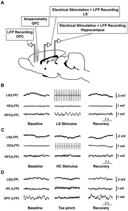
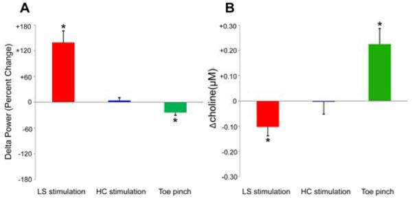
In the current study, we used simultaneous electrophysiology and enzyme-based amperometry in a rat model, and found a decrease in choline, along with slow wave activity in orbital frontal cortex during lateral septal stimulation in the absence of seizures. In contrast, the choline signal and local field potential in frontal cortex had no significant changes when stimulating the hippocampus, but showed increased choline and decreased slow wave activity with an arousal stimulus produced by toe pinch.
These findings indicate that the activation of subcortical inhibitory structures (such as lateral septum) can depress subcortical cholinergic arousal. This mechanism may play an important role in large-scale transitions of cortical activity in focal seizures, as well as in normal cortical function.
在正常睡眠和认知以及癫痫等病理状态下,皮质网络会在活动增加或减少的状态之间发生大规模转换。我们之前发现,大鼠海马局部发作会引起皮质下结构(包括外侧隔区)的神经元放电和脑血流增加,同时伴有类似于慢波睡眠的额皮质慢波振荡。此外,在没有发作的情况下刺激外侧隔区会导致皮质去激活和慢波振荡。
我们假设外侧隔区的激活可能会间接导致新皮质失活,可能是通过皮质下唤醒受损。但是,皮质下刺激如何引起额皮质的慢波活动?在抑制性投射核兴奋期间,皮质中的觉醒神经递质水平(例如乙酰胆碱)如何变化?
在目前的研究中,我们使用大鼠模型进行了同时电生理学和基于酶的安培测量,发现外侧隔区刺激在没有发作的情况下,眶额皮质中的胆碱减少,同时出现慢波活动。相比之下,刺激海马体时,额皮质中的胆碱信号和局部场电位没有明显变化,但当用脚趾夹产生觉醒刺激时,胆碱增加,慢波活动减少。
这些发现表明,皮质下抑制性结构(如外侧隔区)的激活可以抑制皮质下胆碱能唤醒。这种机制可能在局灶性发作中皮质活动的大规模转换以及正常皮质功能中发挥重要作用。