Toledo Jon B, Korff Ané, Shaw Leslie M, Trojanowski John Q, Zhang Jing
Pathology & Laboratory Medicine, Institute on Aging, Center for Neurodegenerative Disease Research, University of Pennsylvania School of Medicine, Philadelphia, PA, USA.
Department of Pathology, University of Washington School of Medicine, HMC Box 359635, 325 9th Avenue, Seattle, WA 98104, USA.
Alzheimers Res Ther. 2014 Jun 23;6(3):36. doi: 10.1186/alzrt266. eCollection 2014.
Alzheimer's disease (AD) is characterized by the deposition of tau and amyloid in the brain. Although the core cerebrospinal fluid (CSF) AD biomarkers amyloid β peptide 1-42 (Aβ1-42), total tau (t-tau) and phosphorylated tau 181 (p-tau181) show good diagnostic sensitivity and specificity, additional biomarkers that can aid in preclinical diagnosis or better track disease progression are needed. Activation of the complement system, a pivotal part of inflammation, occurs at very early stages in the AD brain. Therefore, CSF levels of complement proteins that could be linked to cognitive and structural changes in AD may have diagnostic and prognostic value.
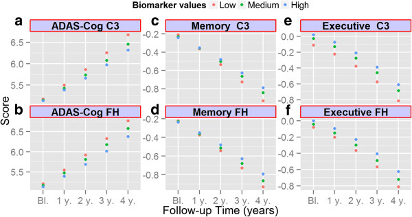
Using xMAP® technology based assays we measured complement 3 (C3) and factor H (FH) in the CSF of 110 controls (CN), 187 mild cognitive impairment (MCI) and 92 AD subjects of the AD Neuroimaging Initiative (ADNI) at baseline. All ADNI participants underwent clinical follow-up at 12 month intervals and MCI subjects had additional visits at 6 and 18 months. The association between CSF biomarkers and different outcome measures were analyzed using Cox proportional hazard models (conversion from MCI to AD), logistic regression models (classification of clinical groups) and mixed-effects models adjusted for age, gender, education, t-tau/Aβ1-42 and APOE ϵ4 presence (baseline and longitudinal association between biomarkers and cognitive scores).
Although no association was found between the complement proteins and clinical diagnosis or cognitive measures, lower levels of C3 (β = -0.12, p = 0.041) and FH (β = -0.075, p = 0.041) were associated with faster cognitive decline in MCI subjects as measured by the AD Assessment Scale-cognitive subscale (ADAS-Cog) test. Furthermore, lower FH levels were associated with larger lateral ventricular volume (p = 0.024), which is indicative of brain atrophy.
Our study confirms a lack of suitability of CSF C3 and FH as diagnostic biomarkers of AD, but points to their modest potential as prognostic biomarkers and therapeutic targets in cognitively impaired patients.
阿尔茨海默病(AD)的特征是大脑中tau蛋白和淀粉样蛋白的沉积。尽管核心脑脊液(CSF)AD生物标志物淀粉样β肽1-42(Aβ1-42)、总tau蛋白(t-tau)和磷酸化tau蛋白181(p-tau181)具有良好的诊断敏感性和特异性,但仍需要其他有助于临床前诊断或更好地跟踪疾病进展的生物标志物。补体系统的激活是炎症的关键部分,在AD大脑的极早期阶段就会发生。因此,与AD认知和结构变化相关的脑脊液补体蛋白水平可能具有诊断和预后价值。
我们使用基于xMAP®技术的检测方法,在基线时测量了阿尔茨海默病神经影像学计划(ADNI)的110名对照(CN)、187名轻度认知障碍(MCI)和92名AD受试者脑脊液中的补体3(C3)和因子H(FH)。所有ADNI参与者每隔12个月接受一次临床随访,MCI受试者在6个月和18个月时进行额外随访。使用Cox比例风险模型(从MCI转化为AD)、逻辑回归模型(临床组分类)以及针对年龄、性别、教育程度、t-tau/Aβ1-42和APOE ε4存在情况进行调整的混合效应模型(生物标志物与认知分数之间的基线和纵向关联)分析脑脊液生物标志物与不同结局指标之间的关联。
尽管未发现补体蛋白与临床诊断或认知指标之间存在关联,但根据阿尔茨海默病评估量表认知子量表(ADAS-Cog)测试,较低的C3水平(β = -0.12,p = 0.041)和FH水平(β = -0.075,p = 0.041)与MCI受试者更快的认知衰退相关。此外,较低的FH水平与更大的侧脑室体积相关(p = 0.024),这表明存在脑萎缩。
我们的研究证实脑脊液C3和FH不适合作为AD的诊断生物标志物,但指出它们在认知受损患者中作为预后生物标志物和治疗靶点具有一定潜力。