Ibragimov Bulat, Prince Jerry L, Murano Emi Z, Woo Jonghye, Stone Maureen, Likar Boštjan, Pernuš Franjo, Vrtovec Tomaž
Faculty of Electrical Engineering, University of Ljubljana, Ljubljana, Slovenia; Department of Electrical and Computer Engineering, Johns Hopkins University, Baltimore, MD, USA.
Department of Electrical and Computer Engineering, Johns Hopkins University, Baltimore, MD, USA.
Med Image Anal. 2015 Feb;20(1):198-207. doi: 10.1016/j.media.2014.11.006. Epub 2014 Nov 23.
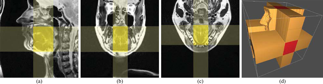
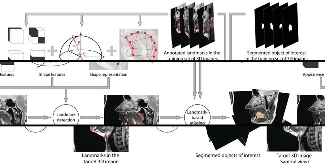
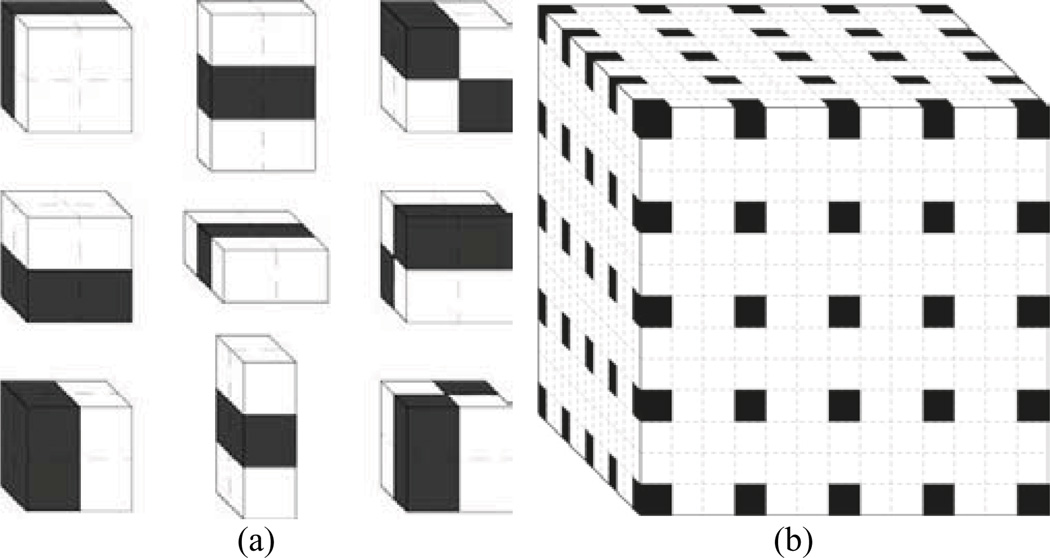
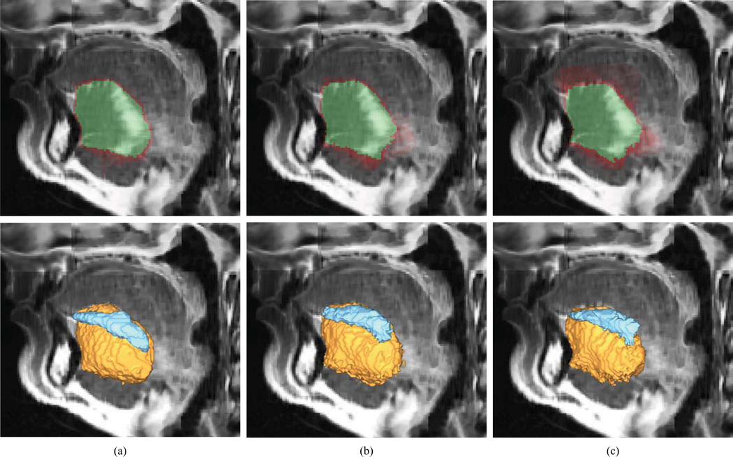
Imaging and quantification of tongue anatomy is helpful in surgical planning, post-operative rehabilitation of tongue cancer patients, and studying of how humans adapt and learn new strategies for breathing, swallowing and speaking to compensate for changes in function caused by disease, medical interventions or aging. In vivo acquisition of high-resolution three-dimensional (3D) magnetic resonance (MR) images with clearly visible tongue muscles is currently not feasible because of breathing and involuntary swallowing motions that occur over lengthy imaging times. However, recent advances in image reconstruction now allow the generation of super-resolution 3D MR images from sets of orthogonal images, acquired at a high in-plane resolution and combined using super-resolution techniques. This paper presents, to the best of our knowledge, the first attempt towards automatic tongue muscle segmentation from MR images. We devised a database of ten super-resolution 3D MR images, in which the genioglossus and inferior longitudinalis tongue muscles were manually segmented and annotated with landmarks. We demonstrate the feasibility of segmenting the muscles of interest automatically by applying the landmark-based game-theoretic framework (GTF), where a landmark detector based on Haar-like features and an optimal assignment-based shape representation were integrated. The obtained segmentation results were validated against an independent manual segmentation performed by a second observer, as well as against B-splines and demons atlasing approaches. The segmentation performance resulted in mean Dice coefficients of 85.3%, 81.8%, 78.8% and 75.8% for the second observer, GTF, B-splines atlasing and demons atlasing, respectively. The obtained level of segmentation accuracy indicates that computerized tongue muscle segmentation may be used in surgical planning and treatment outcome analysis of tongue cancer patients, and in studies of normal subjects and subjects with speech and swallowing problems.
舌部解剖结构的成像和量化有助于手术规划、舌癌患者的术后康复,以及研究人类如何适应和学习新的呼吸、吞咽和说话策略,以补偿疾病、医疗干预或衰老引起的功能变化。由于在长时间成像过程中会出现呼吸和不自主吞咽运动,目前在活体上获取具有清晰可见舌肌的高分辨率三维(3D)磁共振(MR)图像是不可行的。然而,图像重建技术的最新进展现在允许从一组正交图像生成超分辨率3D MR图像,这些图像以高平面分辨率采集,并使用超分辨率技术进行组合。据我们所知,本文首次尝试从MR图像中自动分割舌肌。我们设计了一个包含十张超分辨率3D MR图像的数据库,其中舌颏舌肌和舌下纵肌被手动分割并用地标进行标注。我们通过应用基于地标的博弈论框架(GTF)证明了自动分割感兴趣肌肉的可行性,该框架集成了基于哈尔样特征的地标检测器和基于最优分配的形状表示。所获得的分割结果与第二位观察者进行的独立手动分割以及B样条和魔鬼图谱方法进行了验证。分割性能方面,第二位观察者、GTF、B样条图谱和魔鬼图谱的平均骰子系数分别为85.3%、81.8%、78.8%和75.8%。所获得的分割精度水平表明,计算机化舌肌分割可用于舌癌患者的手术规划和治疗结果分析,以及正常受试者和有言语及吞咽问题受试者的研究。