Shen Yongchun, Wan Chun, Tian Panwen, Wu Yanqiu, Li Xiaoou, Yang Ting, An Jing, Wang Tao, Chen Lei, Wen Fuqiang
From the Department of Respiratory and Critical Care Medicine, West China Hospital of Sichuan University and Division of Pulmonary Diseases, State Key Laboratory of Biotherapy of China, Chengdu 610041, China.
Medicine (Baltimore). 2014 Dec;93(27):e256. doi: 10.1097/MD.0000000000000256.
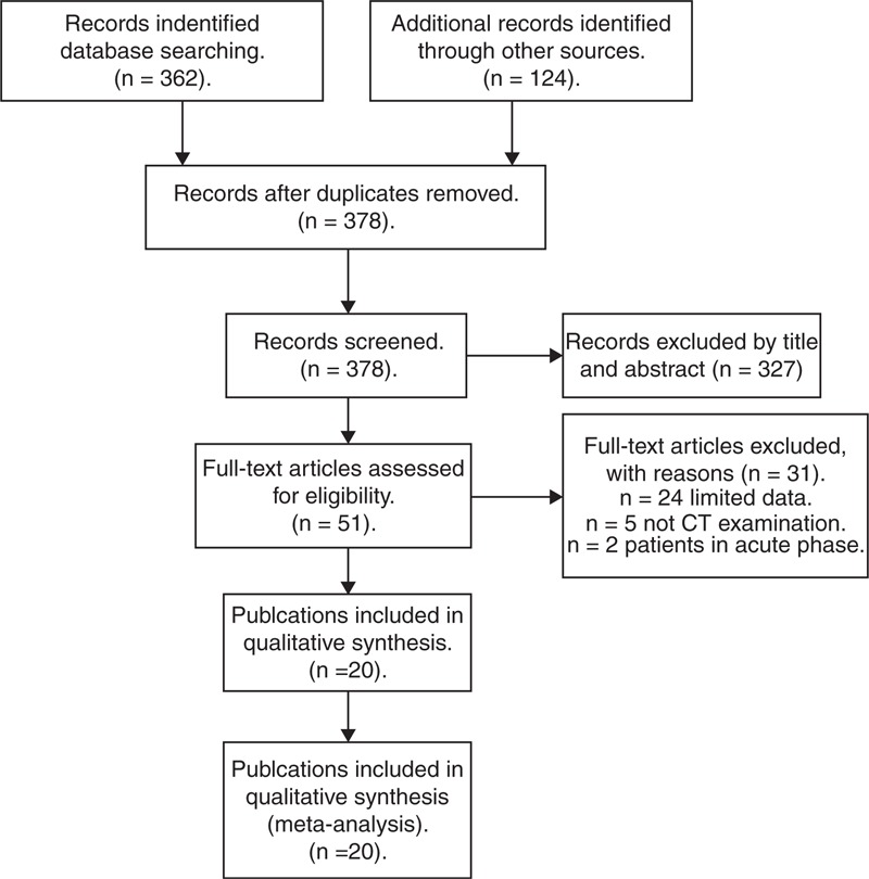
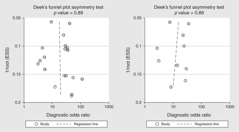
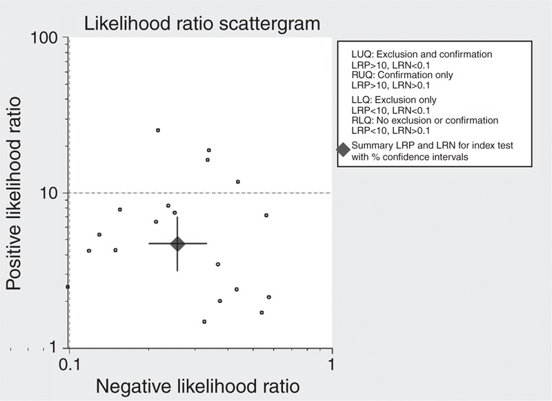
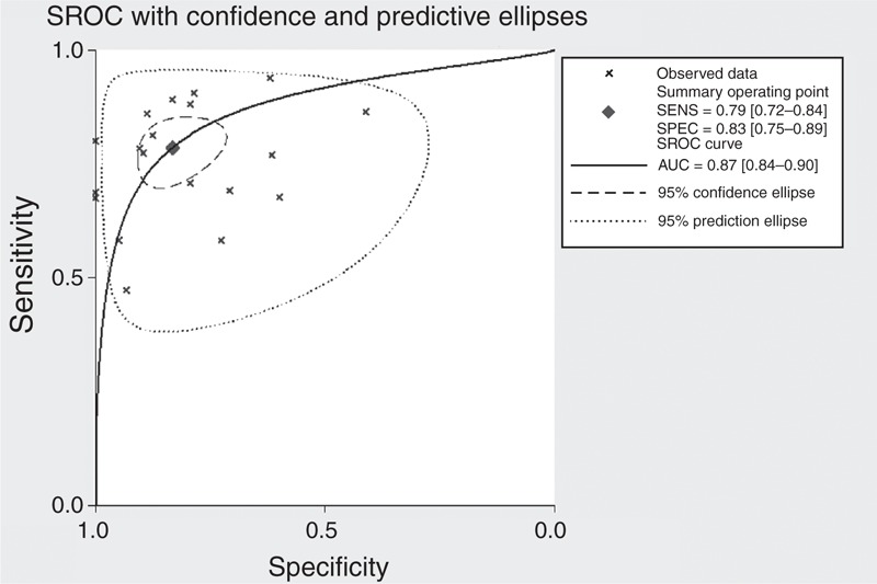
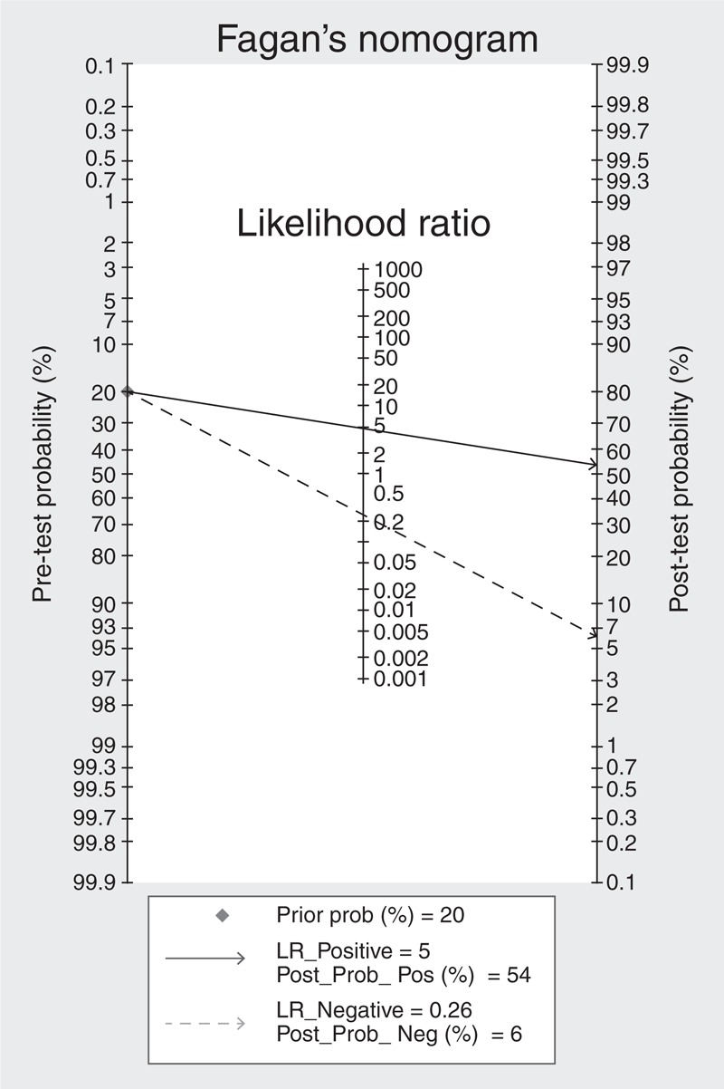
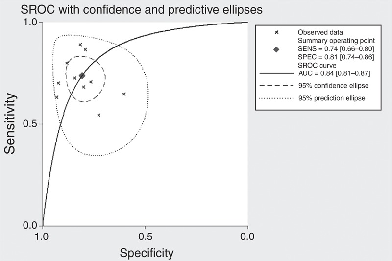
To summarize the performance of CT-based main pulmonary artery diameter or pulmonary artery to aorta ratio (PA:A ratio) measurement in detection of pulmonary hypertension by a systematic review and meta-analysis. A comprehensive literature search was performed to identify studies determining diagnostic accuracy of main pulmonary artery diameter or PA:A ratio measurement for pulmonary hypertension. The Quality Assessment of Diagnostic Accuracy Studies tool was used to assess the quality of the included studies. A bivariate random-effects model was used to pool sensitivity, specificity, positive/negative likelihood ratio (PLR/NLR), and diagnostic odds ratio (DOR). Summary receiver operating characteristic (SROC) curves and area under the curve (AUC) were used to summarize overall diagnostic performance. This meta-analysis included 20 publications involving 2134 subjects. Summary estimates for main pulmonary artery diameter measurement in the diagnosis of pulmonary hypertension were as follows: sensitivity, 0.79 (95% CI 0.72-0.84); specificity, 0.83 (95% CI 0.75-0.89); PLR, 4.68 (95% CI 3.13-6.99); NLR, 0.26 (95% CI 0.20-0.33); DOR, 18.13 (95% CI 10.87-30.24); and AUC 0.87. The corresponding summary performance estimates for using the PA:A ratio were as follows: sensitivity, 0.74 (95% CI 0.66-0.80); specificity, 0.81 (95% CI 0.74-0.86); PLR, 3.83 (95% CI, 2.70-5.43); NLR, 0.33 (95% CI 0.24-0.44); DOR, 11.77 (95% CI 6.60-21.00); and AUC 0.84. Both main pulmonary artery diameter and PA:A ratio are helpful for diagnosing pulmonary hypertension. Nevertheless, the results of pulmonary artery measurement should be interpreted in parallel with the results of traditional tests such as echocardiography.
通过系统评价和荟萃分析总结基于CT的主肺动脉直径或肺动脉与主动脉比值(PA:A比值)测量在检测肺动脉高压中的表现。进行了全面的文献检索,以确定有关主肺动脉直径或PA:A比值测量对肺动脉高压诊断准确性的研究。使用诊断准确性研究质量评估工具评估纳入研究的质量。采用双变量随机效应模型汇总敏感性、特异性、阳性/阴性似然比(PLR/NLR)和诊断比值比(DOR)。采用汇总受试者工作特征(SROC)曲线和曲线下面积(AUC)总结总体诊断性能。该荟萃分析纳入了20篇涉及2134名受试者的出版物。主肺动脉直径测量诊断肺动脉高压的汇总估计如下:敏感性为0.79(95%CI 0.72 - 0.84);特异性为0.83(95%CI 0.75 - 0.89);PLR为4.68(95%CI 3.13 - 6.99);NLR为0.26(95%CI 0.20 - 0.33);DOR为18.13(95%CI 10.87 - 30.24);AUC为0.87。使用PA:A比值的相应汇总性能估计如下:敏感性为0.74(95%CI 0.66 - 0.80);特异性为0.81(95%CI 0.74 - 0.86);PLR为3.83(95%CI 2.70 - 5.43);NLR为0.33(95%CI 0.24 - 0.44);DOR为11.77(95%CI 6.60 - 21.00);AUC为0.84。主肺动脉直径和PA:A比值均有助于诊断肺动脉高压。然而,肺动脉测量结果应与传统检查如超声心动图的结果一并解读。