Fan Xiaobing, Mustafi Devkumar, Markiewicz Erica, Zamora Marta, Vosicky James, Leinroth Abby, Mueller Jeffrey, Macleod Kay, Conzen Suzanne D, Karczmar Gregory S
Department of Radiology, MC2026, The University of Chicago, 5841 S. Maryland Avenue, Chicago, IL, 60637, USA.
Department of Pathology, MC6101, The University of Chicago, 5841 S. Maryland Avenue, Chicago, IL, 60637, USA.
Breast Cancer Res. 2014 Dec 16;16(6):495. doi: 10.1186/s13058-014-0495-6.
Previous work from this laboratory demonstrated that magnetic resonance imaging (MRI) detects early murine mammary cancers and reliably differentiates between in situ and invasive cancer. Based on this previous work, we used MRI to study initiation and progression of murine mammary cancer, and monitor the transition from the in situ to the invasive phase.
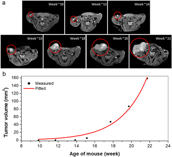
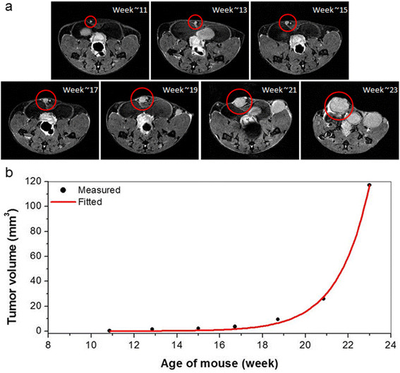
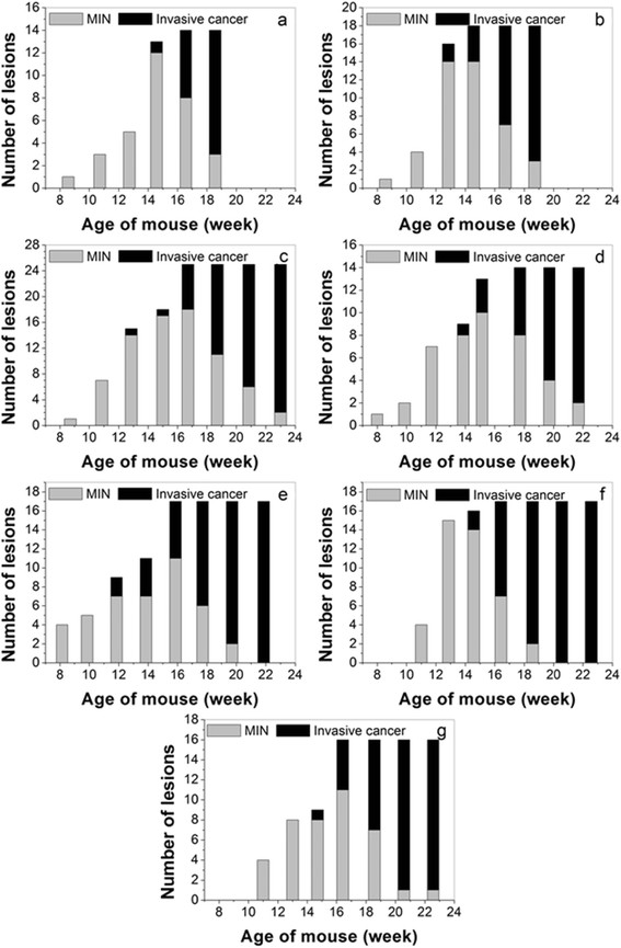
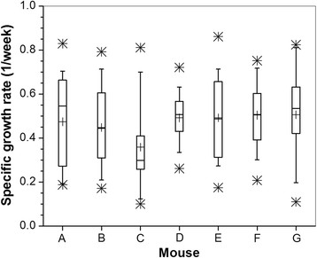
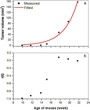
In total, seven female C3(1) SV40 Tag mice were imaged every two weeks between the ages of 8 to 23 weeks. Lesions were identified on T2-weighted images acquired at 9.4 Tesla based on their morphology and growth rates. Lesions were traced manually on MR images of each slice. Volume of each lesion was calculated by adding measurements from individual slices. Plots of lesion volume versus time were analyzed to obtain the specific growth rate (SGR). The time at which in situ cancers (referred to as 'mammary intraepithelial neoplasia (MIN)') and invasive cancers were first detected; and the time at which in situ cancers became invasive were recorded.
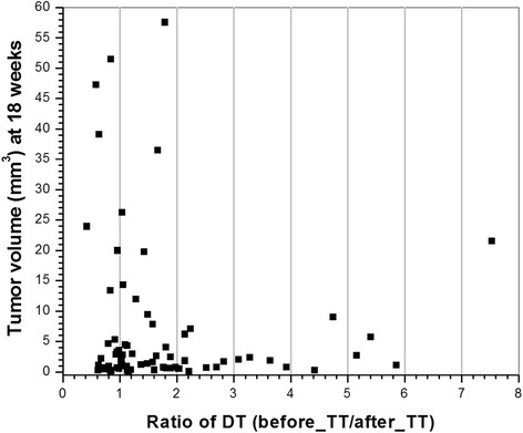
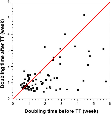
A total of 121 cancers (14 to 25 per mouse) were identified in seven mice. On average the MIN lesions and invasive cancers were first detected when mice were 13 and 18 weeks old, respectively. The average SGR was 0.47 ± 0.18 week(-1) and there were no differences (P >0.05) between mice. 74 lesions had significantly different tumor growth rates before and after ~17 weeks of age; with average doubling times (DT) of 1.88 and 1.27 weeks, respectively. The average DT was significantly shorter (P <0.0001) after 17 weeks of age. However, the DT for some cancers was longer after 17 weeks of age, and about 10% of the cancers detected did not progress to the invasive stage.
A wide range of growth rates were observed in SV40 mammary cancers. Most cancers transitioned to a more aggressive phenotype at approximately 17 weeks of age, but some cancers became less aggressive. The results suggest that the biology of mammary cancers is extremely heterogeneous. This work is a first step towards use of MRI to improve understanding of factors that control and/or signal the development of aggressive breast cancer.
本实验室之前的研究表明,磁共振成像(MRI)能够检测出早期小鼠乳腺癌,并可靠地区分原位癌和浸润性癌。基于此前的研究工作,我们利用MRI来研究小鼠乳腺癌的发生和发展过程,并监测从原位癌到浸润性癌的转变。
总共7只雌性C3(1) SV40 Tag小鼠在8至23周龄期间每两周进行一次成像。根据在9.4特斯拉下采集的T2加权图像上病变的形态和生长速率来识别病变。在每一层的MR图像上手动追踪病变。通过将各层的测量值相加来计算每个病变的体积。分析病变体积随时间的变化图以获得特定生长速率(SGR)。记录首次检测到原位癌(称为“乳腺上皮内瘤变(MIN)”)和浸润性癌的时间;以及原位癌转变为浸润性癌的时间。
在7只小鼠中总共识别出121个癌症(每只小鼠14至25个)。平均而言,MIN病变和浸润性癌分别在小鼠13周龄和18周龄时首次被检测到。平均SGR为0.47±0.18周-1,小鼠之间无差异(P>0.05)。74个病变在约17周龄前后的肿瘤生长速率有显著差异;平均倍增时间(DT)分别为1.88周和1.27周。17周龄后平均DT显著缩短(P<0.0001)。然而,一些癌症在17周龄后的DT更长,并且检测到的癌症中约10%未进展到浸润阶段。
在SV40乳腺肿瘤中观察到广泛的生长速率。大多数癌症在约17周龄时转变为更具侵袭性的表型,但一些癌症的侵袭性降低。结果表明乳腺癌的生物学特性极为异质性。这项工作是朝着利用MRI来更好地理解控制和/或指示侵袭性乳腺癌发展的因素迈出的第一步。