Alvarez Karl, de Andrés María C, Takahashi Atsushi, Oreffo Richard O C
Bone and Joint Research Group, Centre for Human Development, Stem Cells and Regeneration Human Development and Health, Institute of Developmental Sciences, University of Southampton, Southampton SO16 6YD, UK.
BMC Musculoskelet Disord. 2014 Dec 15;15:431. doi: 10.1186/1471-2474-15-431.
Cartilage is an avascular and aneural tissue. Chondrocytes thrive in this restricted environment of low oxygen tension and poor nutrient availability which has led to suggestions that hypoxia may be a protective mechanism against the development of osteoarthritis (OA). There is also a growing body of evidence to support the role of epigenetic factors in the pathogenesis of OA. However, few studies have investigated the epigenetic-OA process within a hypoxic environment. The current study has investigated the effects of hypoxia on gene expression and DNA methylation of anabolic and catabolic genes involved in the pathogenesis of OA.
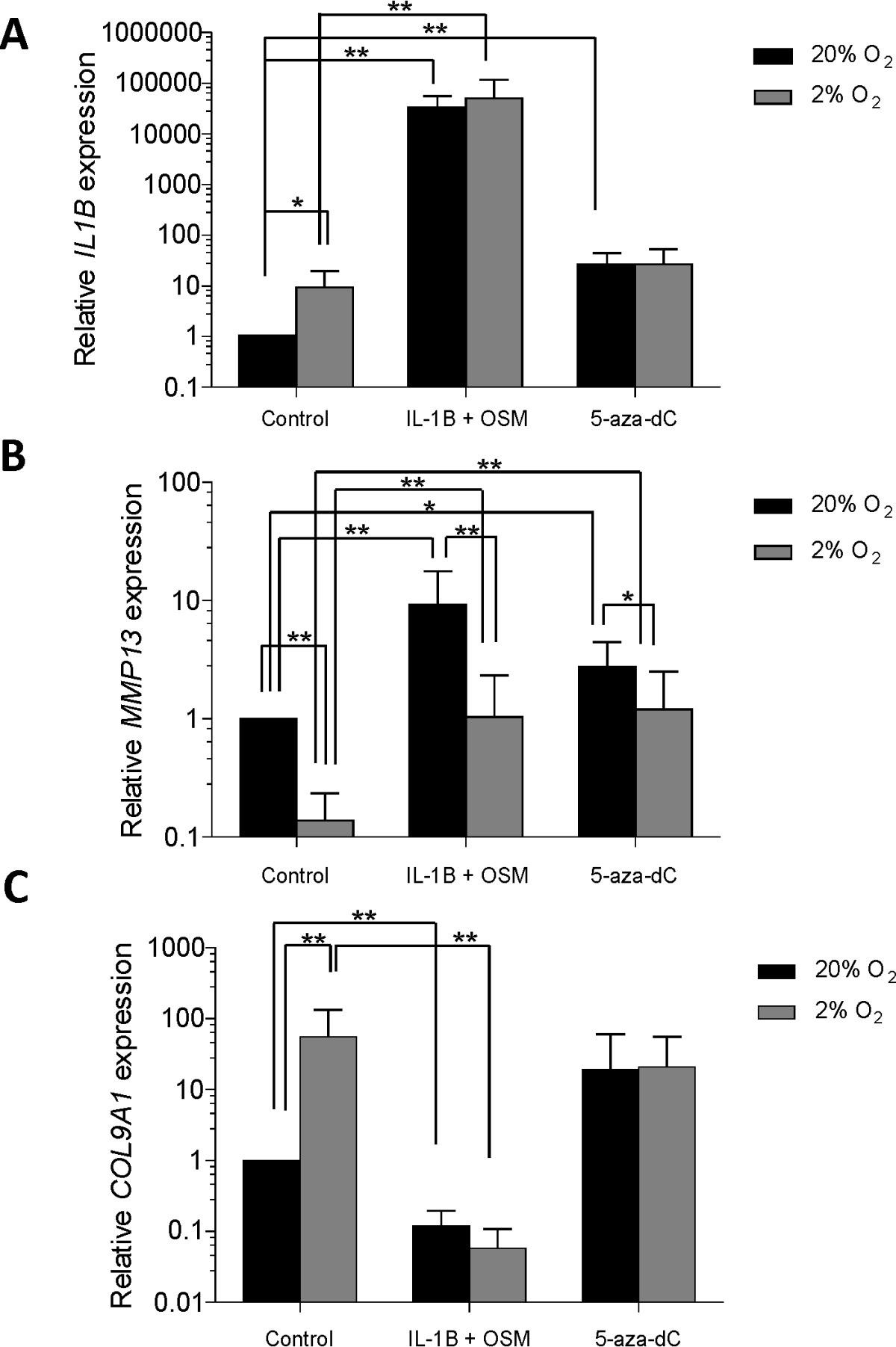
Chondrocytes extracted from OA femoral heads were incubated in normoxia and hypoxia (20% and 2% oxygen concentrations respectively). Interleukin 1-beta (IL-1β) plus oncostatin M (OSM), 5-azadeoxycytidine (5-aza-dC) or media alone (control) were added twice weekly to the incubated samples. After 5 weeks, levels of Collagen type IX (COL9A1), IL1B, and matrix metalloproteinase-13 (MMP13) gene expression were measured using SYBR Green-based qRT-PCR and were correlated with methylation status analysed by pyrosequencing methodology.
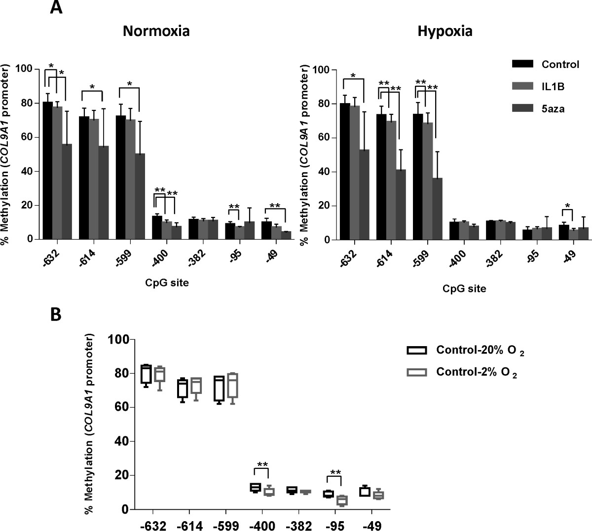
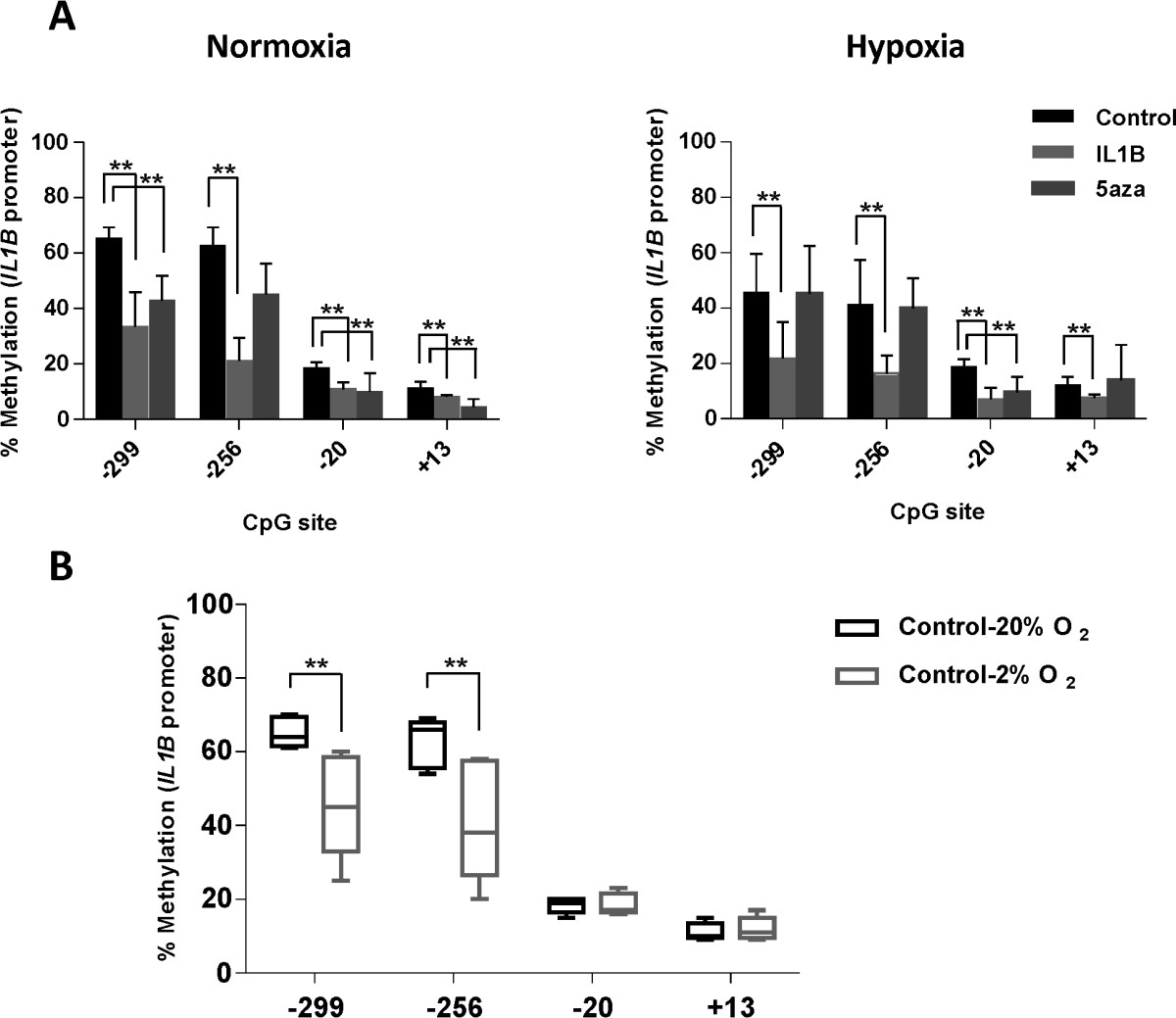
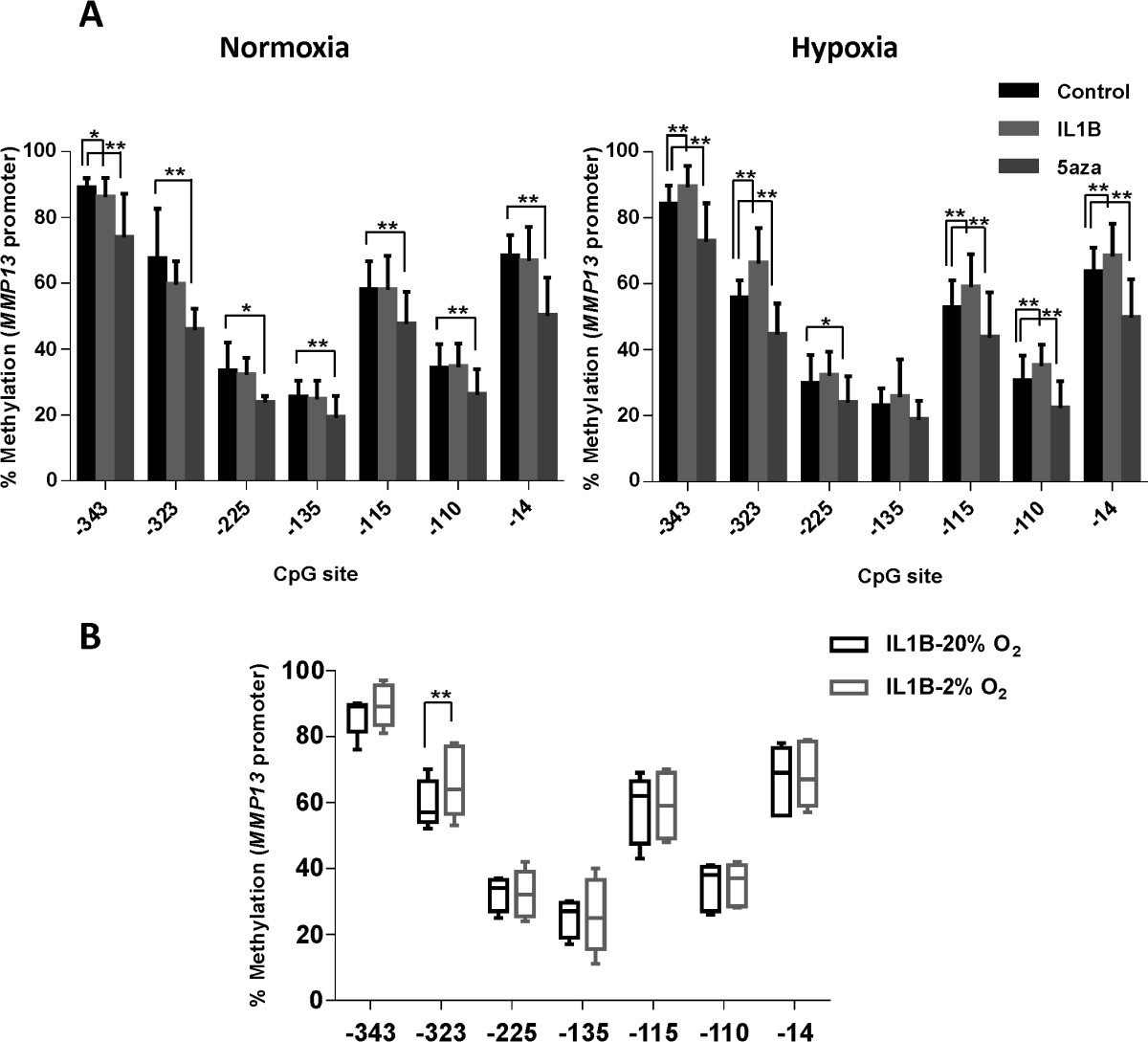
Hypoxia resulted in a >50-fold and >10-fold increase in relative expression of COL9A1 and IL1B respectively. This was inversely correlated to the DNA methylation status of these genes. Expression of MMP13 was reduced at 2% oxygen tension in control cells. Relative expression of MMP13 increased in cells stimulated with IL-1β and 5-aza-dC in normoxic conditions, and this effect was eliminated at low oxygen tension although no correlation with methylation status was observed.
These findings demonstrate a role for hypoxia in the regulation of anabolic and catabolic gene expression and the influence of changes in DNA methylation. These results further support the role of epigenetics in OA and, critically, highlight the complex relationship between the physiological environment of cartilaginous cells and the osteoarthritic process with implications for therapeutic intervention and our understanding of OA pathophysiology.
软骨是一种无血管和神经的组织。软骨细胞在这种低氧张力和营养供应不足的受限环境中茁壮成长,这引发了一种观点,即缺氧可能是一种预防骨关节炎(OA)发展的保护机制。也有越来越多的证据支持表观遗传因素在OA发病机制中的作用。然而,很少有研究在缺氧环境中研究表观遗传 - OA过程。本研究调查了缺氧对参与OA发病机制的合成代谢和分解代谢基因的基因表达及DNA甲基化的影响。
从OA股骨头中提取的软骨细胞分别在常氧和缺氧(氧浓度分别为20%和2%)条件下培养。每周两次向培养的样本中添加白细胞介素1 - β(IL - 1β)加制瘤素M(OSM)、5 - 氮杂脱氧胞苷(5 - aza - dC)或仅添加培养基(对照)。5周后,使用基于SYBR Green的qRT - PCR测量IX型胶原蛋白(COL9A1)、IL1B和基质金属蛋白酶 - 13(MMP13)基因的表达水平,并与通过焦磷酸测序法分析的甲基化状态相关联。
缺氧分别导致COL9A1和IL1B的相对表达增加>50倍和>10倍。这与这些基因的DNA甲基化状态呈负相关。在对照细胞中,氧张力为2%时MMP13的表达降低。在常氧条件下,用IL - 1β和5 - aza - dC刺激的细胞中MMP13的相对表达增加,而在低氧张力下这种效应消失,尽管未观察到与甲基化状态的相关性。
这些发现证明了缺氧在调节合成代谢和分解代谢基因表达以及DNA甲基化变化影响方面的作用。这些结果进一步支持了表观遗传学在OA中的作用,并且至关重要的是,突出了软骨细胞生理环境与骨关节炎过程之间的复杂关系,这对治疗干预以及我们对OA病理生理学的理解具有重要意义。