• 文献检索
  • 文档翻译
  • 深度研究
  • 学术资讯
  • Suppr Zotero 插件Zotero 插件
  • 邀请有礼
  • 套餐&价格
  • 历史记录
应用&插件
Suppr Zotero 插件Zotero 插件浏览器插件Mac 客户端Windows 客户端微信小程序
定价
高级版会员购买积分包购买API积分包
服务
文献检索文档翻译深度研究API 文档MCP 服务
关于我们
关于 Suppr公司介绍联系我们用户协议隐私条款
关注我们

Suppr 超能文献

核心技术专利:CN118964589B侵权必究
粤ICP备2023148730 号-1Suppr @ 2026

文献检索

告别复杂PubMed语法,用中文像聊天一样搜索,搜遍4000万医学文献。AI智能推荐,让科研检索更轻松。

立即免费搜索

文件翻译

保留排版,准确专业,支持PDF/Word/PPT等文件格式,支持 12+语言互译。

免费翻译文档

深度研究

AI帮你快速写综述,25分钟生成高质量综述,智能提取关键信息,辅助科研写作。

立即免费体验

扩散加权围产期尸检磁共振成像作为死后间隔时间的标志物

Diffusion-weighted perinatal postmortem magnetic resonance imaging as a marker of postmortem interval.

作者信息

Arthurs Owen J, Price Gemma C, Carmichael David W, Jones Rod, Norman Wendy, Taylor Andrew M, Sebire Neil J

机构信息

Department of Radiology, Great Ormond Street Hospital for Children NHS Foundation Trust, London, WC1N 3JH, UK,

出版信息

Eur Radiol. 2015 May;25(5):1399-406. doi: 10.1007/s00330-014-3525-y. Epub 2014 Dec 18.

DOI:10.1007/s00330-014-3525-y
PMID:25519976
原文链接:https://pmc.ncbi.nlm.nih.gov/articles/PMC4392167/
Abstract

OBJECTIVE

To evaluate perinatal body organ apparent diffusion coefficient (ADC) values at postmortem magnetic resonance imaging (PMMR) in order to evaluate postmortem changes.

METHODS

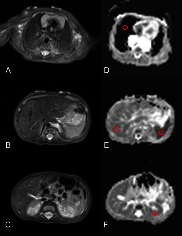
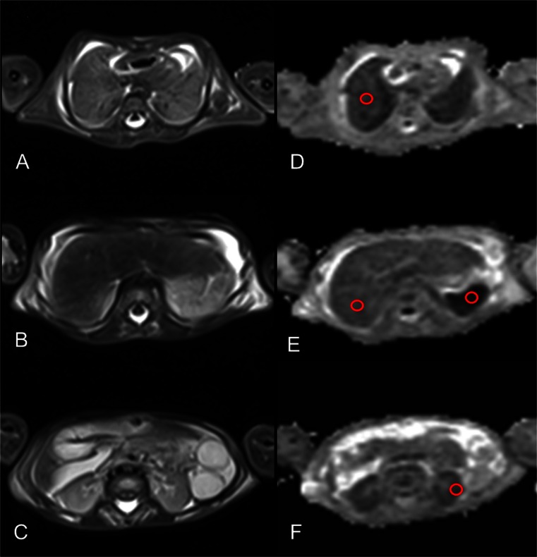
Postmortem diffusion-weighted imaging (DWI) of the thorax and abdomen were performed with diffusion gradient values b = 0, 500, and 1000 s/mm(2) on 15 foetal and childhood cases (mean 33.3 ± 7.8 weeks gestation) compared to 44 live infants (mean age 75.5 ± 53.4 days). Mean ADC values were calculated from regions of interest (ROIs) for the lungs, liver, spleen and renal cortex, compared to normative live infantile body ADC values of similar gestational age.

RESULTS

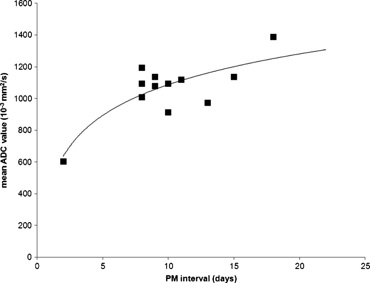
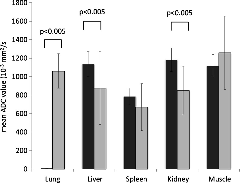
Mean ADC values were significantly lower in postmortem cases than in normal controls for liver (0.88 10(-3) mm(2)/s ± SD 0.39 vs. 1.13 ± 0.13; p < 0.05) and renal cortex (0.85 ± 0.26 vs. 1.19 ± 0.13; p < 0.05) but not spleen or muscle. Mean lung ADC values were significantly higher than normal controls (1.06 ± 0.18 vs. 0 ± 0; p < 0.001), and there was a significant correlation between postmortem interval and lung ADC (R(2) = 0.55).

CONCLUSION

Lung PMMR ADC values are related to postmortem interval, making them a potential marker of time since death. Further research is needed to understand the organ-specific changes which occur in the postmortem period.

KEY POINTS

• Liver and spleen PM ADC values were lower than controls. • Lung ADC changes correlate with PM interval. • These findings may be useful in medicolegal cases.

摘要

目的

评估围产期尸体磁共振成像(PMMR)中身体器官的表观扩散系数(ADC)值,以评估死后变化。

方法

对15例胎儿和儿童病例(平均孕周33.3±7.8周)进行胸部和腹部的死后扩散加权成像(DWI),扩散梯度值b = 0、500和1000 s/mm²,与44例活产婴儿(平均年龄75.5±53.4天)进行比较。从感兴趣区域(ROI)计算肺、肝、脾和肾皮质的平均ADC值,并与相似孕周的正常活产婴儿身体ADC值进行比较。

结果

死后病例中,肝脏(0.88×10⁻³mm²/s±标准差0.39 vs. 1.13±0.13;p < 0.05)和肾皮质(0.85±0.26 vs. 1.19±0.13;p < 0.05)的平均ADC值显著低于正常对照组,但脾脏或肌肉无此情况。肺的平均ADC值显著高于正常对照组(1.06±0.18 vs. 0±0;p < 0.001),且死后间隔与肺ADC之间存在显著相关性(R² = 0.55)。

结论

肺PMMR ADC值与死后间隔相关,使其成为死亡时间的潜在标志物。需要进一步研究以了解死后期间发生的器官特异性变化。

关键点

•肝脏和脾脏的PM ADC值低于对照组。•肺ADC变化与PM间隔相关。•这些发现可能在法医学案件中有用。

https://cdn.ncbi.nlm.nih.gov/pmc/blobs/410c/4392167/774b98f4ee56/330_2014_3525_Fig4_HTML.jpg
https://cdn.ncbi.nlm.nih.gov/pmc/blobs/410c/4392167/7f9fb273a228/330_2014_3525_Fig1_HTML.jpg
https://cdn.ncbi.nlm.nih.gov/pmc/blobs/410c/4392167/52f208b0b320/330_2014_3525_Fig2_HTML.jpg
https://cdn.ncbi.nlm.nih.gov/pmc/blobs/410c/4392167/f13dd8862caa/330_2014_3525_Fig3_HTML.jpg
https://cdn.ncbi.nlm.nih.gov/pmc/blobs/410c/4392167/774b98f4ee56/330_2014_3525_Fig4_HTML.jpg

相似文献

1
Diffusion-weighted perinatal postmortem magnetic resonance imaging as a marker of postmortem interval.扩散加权围产期尸检磁共振成像作为死后间隔时间的标志物
Eur Radiol. 2015 May;25(5):1399-406. doi: 10.1007/s00330-014-3525-y. Epub 2014 Dec 18.
2
The use of whole body diffusion-weighted post-mortem magnetic resonance imaging in timing of perinatal deaths.全身扩散加权尸检磁共振成像在围产期死亡时间判定中的应用。
Int J Legal Med. 2018 Nov;132(6):1735-1741. doi: 10.1007/s00414-018-1906-5. Epub 2018 Jul 28.
3
Postmortem MR diffusion-weighted imaging of the liver: time-behavior of the hepatic apparent diffusion coefficient in the early death interval.肝脏的尸检磁共振扩散加权成像:早期死亡间隔期肝脏表观扩散系数的时间变化情况
Int J Legal Med. 2018 Jan;132(1):263-268. doi: 10.1007/s00414-017-1685-4. Epub 2017 Sep 14.
4
Diffusion-weighted post-mortem magnetic resonance imaging of the human fetal brain in situ.原位人类胎儿脑的扩散加权尸检磁共振成像
Eur J Radiol. 2016 Jun;85(6):1167-73. doi: 10.1016/j.ejrad.2016.03.024. Epub 2016 Mar 24.
5
Postmortem Diffusion-Weighted Magnetic Resonance Imaging of the Brain in Perinatal Death: An Animal Control Study to Detect the Influence of Postmortem Interval.围生期死亡后脑弥散加权磁共振成像的死后研究:一项动物对照研究,旨在检测死后间隔时间的影响。
Top Magn Reson Imaging. 2022 Oct 1;31(5):43-50. doi: 10.1097/RMR.0000000000000299.
6
Prognostic value of diffusion-weighted magnetic resonance imaging of brain in fetal growth restriction: results of prospective multicenter study.胎儿生长受限的脑弥散加权磁共振成像的预后价值:前瞻性多中心研究结果。
Ultrasound Obstet Gynecol. 2020 Dec;56(6):893-900. doi: 10.1002/uog.21926.
7
Which organ should be considered a reference in diffusion weighted imaging of the abdomen?: The reproducibility of ADC measurements of the spleen and the renal cortex on a 1.5T MR.在腹部扩散加权成像中,哪个器官应被视为参考器官?:1.5T磁共振成像上脾脏和肾皮质表观扩散系数(ADC)测量的可重复性
Adv Clin Exp Med. 2017 Aug;26(5):811-816. doi: 10.17219/acem/60877.
8
Diffusion-weighted imaging (DWI) of the spleen in patients with liver cirrhosis and portal hypertension.肝硬化和门静脉高压症患者脾脏的弥散加权成像(DWI)。
Magn Reson Imaging. 2013 Sep;31(7):1092-6. doi: 10.1016/j.mri.2013.01.003. Epub 2013 Jun 2.
9
Measurement of apparent diffusion coefficient (ADC) in fetal organs and placenta using 3 Tesla magnetic resonance imaging (MRI) across gestational ages.使用 3 特斯拉磁共振成像(MRI)测量胎儿器官和胎盘的表观扩散系数(ADC)在各个妊娠期的变化。
Sci Rep. 2024 Oct 11;14(1):23811. doi: 10.1038/s41598-024-73902-x.
10
Postmortem Magnetic Resonance Imaging of In Utero Fetuses and Its Relationship With Postmortem Interval: A Multi-Organ Observational Study on Reduced Fetuses of Complicated Multiple Pregnancies.宫内胎儿的尸检磁共振成像及其与尸检间隔的关系:对复杂多胎妊娠中小于胎龄胎儿的多器官观察研究
J Magn Reson Imaging. 2023 Mar;57(3):952-961. doi: 10.1002/jmri.28388. Epub 2022 Aug 31.

引用本文的文献

1
Postmortem Temporal Changes in Liver and Spleen Stiffness: Evaluation with Shear Wave Elastography in a Rat Model.肝脏和脾脏硬度的死后时间变化:大鼠模型中剪切波弹性成像评估
Diagnostics (Basel). 2025 Apr 10;15(8):958. doi: 10.3390/diagnostics15080958.
2
The Role and Place of Thanatoradiological Studies in the Pathological Examination of Fetuses and Newborns.Thanatoradiological 研究在胎儿和新生儿病理检查中的作用和地位。
Bull Exp Biol Med. 2022 Oct;173(6):691-705. doi: 10.1007/s10517-022-05615-y. Epub 2022 Nov 4.
3
Effect of Post-mortem Interval and Perfusion on the Biophysical Properties of Liver Tissue Investigated Longitudinally by MRE and DWI.

本文引用的文献

1
Indications, advantages and limitations of perinatal postmortem imaging in clinical practice.围产期尸体剖验成像在临床实践中的适应证、优势及局限性
Pediatr Radiol. 2015 Apr;45(4):491-500. doi: 10.1007/s00247-014-3165-z. Epub 2014 Oct 2.
2
Foetal radiography for suspected skeletal dysplasia: technique, normal appearances, diagnostic approach.疑似骨骼发育不良的胎儿X线摄影:技术、正常表现及诊断方法
Pediatr Radiol. 2015 Apr;45(4):536-48. doi: 10.1007/s00247-014-3130-x. Epub 2014 Aug 31.
3
Apparent diffusion coefficient and beyond: what diffusion MR imaging can tell us about tissue structure.
尸检间隔和灌注对通过磁共振弹性成像(MRE)和扩散加权成像(DWI)纵向研究的肝组织生物物理特性的影响。
Front Physiol. 2021 Aug 3;12:696304. doi: 10.3389/fphys.2021.696304. eCollection 2021.
4
Parameters for estimating the time of death at perinatal autopsy of stillborn fetuses: a systematic review.死产胎儿围产期尸检时死亡时间估计的参数:一项系统评价
Int J Legal Med. 2019 Mar;133(2):483-489. doi: 10.1007/s00414-019-01999-1. Epub 2019 Jan 8.
5
High resolution isotropic diffusion imaging in post-mortem neonates: a feasibility study.死后新生儿的高分辨率各向同性扩散成像:一项可行性研究。
Br J Radiol. 2018 Dec;91(1092):20180319. doi: 10.1259/bjr.20180319. Epub 2018 Aug 30.
6
Postmortem MR diffusion-weighted imaging of the liver: time-behavior of the hepatic apparent diffusion coefficient in the early death interval.肝脏的尸检磁共振扩散加权成像:早期死亡间隔期肝脏表观扩散系数的时间变化情况
Int J Legal Med. 2018 Jan;132(1):263-268. doi: 10.1007/s00414-017-1685-4. Epub 2017 Sep 14.
7
Current issues in postmortem imaging of perinatal and forensic childhood deaths.围产期和法医儿童死亡尸体解剖成像的当前问题
Forensic Sci Med Pathol. 2017 Mar;13(1):58-66. doi: 10.1007/s12024-016-9821-x. Epub 2017 Jan 12.
8
The radiodensity of cerebrospinal fluid and vitreous humor as indicator of the time since death.脑脊液和玻璃体液的放射密度作为死亡时间的指标。
Forensic Sci Med Pathol. 2016 Sep;12(3):248-56. doi: 10.1007/s12024-016-9778-9. Epub 2016 Apr 27.
9
Perinatal and paediatric post-mortem magnetic resonance imaging (PMMR): sequences and technique.围产期和儿科尸检磁共振成像(PMMR):序列与技术
Br J Radiol. 2016 Jun;89(1062):20151028. doi: 10.1259/bjr.20151028. Epub 2016 Mar 15.
10
Pleural fluid accumulation detectable on paediatric post-mortem imaging: a possible marker of interval since death?小儿尸检影像上可检测到的胸腔积液:是否为死亡间隔时间的潜在标志物?
Int J Legal Med. 2016 Jul;130(4):1003-1010. doi: 10.1007/s00414-016-1320-9. Epub 2016 Feb 4.
表观扩散系数及其他:扩散磁共振成像能告诉我们关于组织结构的哪些信息。
Radiology. 2013 Aug;268(2):318-22. doi: 10.1148/radiol.13130420.
4
Post-mortem MRI versus conventional autopsy in fetuses and children: a prospective validation study.胎儿和儿童死后 MRI 与传统尸检的比较:一项前瞻性验证研究。
Lancet. 2013 Jul 20;382(9888):223-33. doi: 10.1016/S0140-6736(13)60134-8. Epub 2013 May 16.
5
Role of diffusion-weighted imaging in differentiating benign and malignant pediatric abdominal tumors.扩散加权成像在鉴别儿童腹部良恶性肿瘤中的作用。
Pediatr Radiol. 2013 Jul;43(7):836-45. doi: 10.1007/s00247-013-2626-0. Epub 2013 May 12.
6
Histological validation of DW-MRI tractography in human postmortem tissue.DW-MRI 示踪技术在人体死后组织中的组织学验证。
Cereb Cortex. 2013 Feb;23(2):442-50. doi: 10.1093/cercor/bhs036. Epub 2012 Feb 17.
7
Post mortem magnetic resonance imaging in the fetus, infant and child: a comparative study with conventional autopsy (MaRIAS Protocol).胎儿、婴儿和儿童死后磁共振成像:与常规尸检的比较研究(MaRIAS 方案)。
BMC Pediatr. 2011 Dec 22;11:120. doi: 10.1186/1471-2431-11-120.
8
Forensic application of postmortem diffusion-weighted and diffusion tensor MR imaging of the human brain in situ.人体原位死后弥散加权和弥散张量磁共振成像的法医应用。
AJNR Am J Neuroradiol. 2011 Sep;32(8):1518-24. doi: 10.3174/ajnr.A2508. Epub 2011 Jun 9.
9
Post-mortem cerebral magnetic resonance imaging T1 and T2 in fetuses, newborns and infants.胎儿、新生儿和婴儿的死后大脑磁共振成像 T1 和 T2
Eur J Radiol. 2012 Mar;81(3):e232-8. doi: 10.1016/j.ejrad.2011.01.105. Epub 2011 Feb 23.
10
Minimally-invasive fetal autopsy using magnetic resonance imaging and percutaneous organ biopsies: clinical value and comparison to conventional autopsy.经磁共振成像和经皮器官活检的微创胎儿尸检:临床价值与传统尸检比较。
Ultrasound Obstet Gynecol. 2011 Mar;37(3):317-23. doi: 10.1002/uog.8844.