Verbeke Stephanie, Richard Elodie, Monceau Elodie, Schmidt Xenia, Rousseau Benoit, Velasco Valerie, Bernard David, Bonnefoi Herve, MacGrogan Gaetan, Iggo Richard D
INSERM U916, Bergonié Cancer Institute, University of Bordeaux, 229 cours de l'Argonne, Bordeaux, 33076, France.
School of Medicine, University of St Andrews, Medical and Biological Sciences Building, North Haugh, St Andrews, KY16 9TF, UK.
Breast Cancer Res. 2014 Dec 20;16(6):504. doi: 10.1186/s13058-014-0504-9.
The cell of origin for estrogen receptor α-positive (ERα+) breast cancer is probably a luminal stem cell in the terminal duct lobular units. To model these cells, we have used the murine myoepithelial layer in the mouse mammary ducts as a scaffold upon which to build a human luminal layer. To prevent squamous metaplasia, a common artifact in genetically-engineered breast cancer models, we sought to limit activation of the epidermal growth factor receptor (EGFR) during in vitro cell culture before grafting the cells.
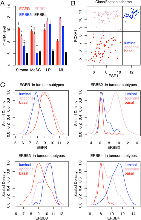
Human reduction mammoplasty cells were grown in vitro in WIT medium. Epidermal growth factor in the medium was replaced with amphiregulin and neuregulin to decrease activation of EGFR and increase activation of EGFR homologs 3 and 4 (ERBB3 and ERBB4). Lentiviral vectors were used to express oncogenic transgenes and fluorescent proteins. Human mammary epithelial cells were mixed with irradiated mouse fibroblasts and Matrigel, then injected through the nipple into the mammary ducts of immunodeficient mice. Engrafted cells were visualized by stereomicroscopy for fluorescent proteins and characterized by histology and immunohistochemistry.
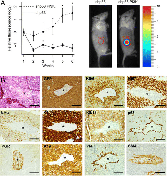
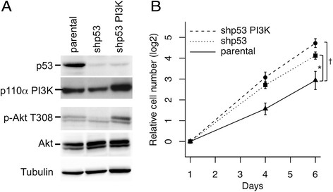
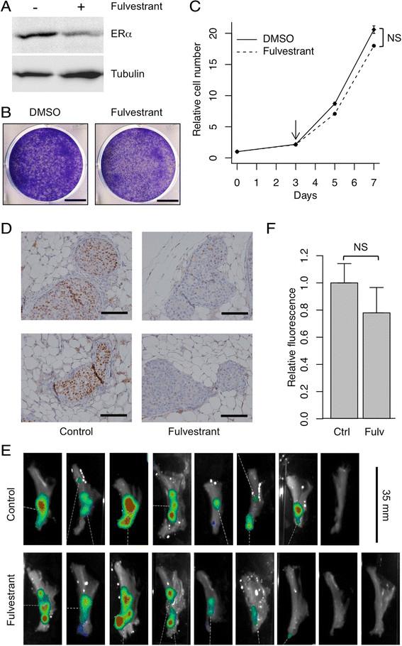
Growth of normal mammary epithelial cells in conditions favoring ERBB3/4 signaling prevented squamous metaplasia in vitro. Normal human cells were quickly lost after intraductal injection, but cells infected with lentiviruses expressing CCND1, MYC, TERT, BMI1 and a short-hairpin RNA targeting TP53 were able to engraft and progressively replace the luminal layer in the mouse mammary ducts, resulting in the formation of an extensive network of humanized ducts. Despite expressing multiple oncogenes, the human cells formed a morphologically normal luminal layer. Expression of a single additional oncogene, PIK3CA-H1047R, converted the cells into invasive cancer cells. The resulting tumors were ERα+, Ki67+ luminal B adenocarcinomas that were resistant to treatment with fulvestrant.
Injection of preneoplastic human mammary epithelial cells into the mammary ducts of immunodeficient mice leads to replacement of the murine luminal layer with morphologically normal human cells. Genetic manipulation of the injected cells makes it possible to study defined steps in the transformation of human mammary epithelial cells in a more physiological environment than has hitherto been possible.
雌激素受体α阳性(ERα+)乳腺癌的起源细胞可能是终末导管小叶单位中的腔面干细胞。为了构建这些细胞的模型,我们利用小鼠乳腺导管中的肌上皮层作为支架,在其上构建人腔面层。为了防止鳞状化生(这是基因工程乳腺癌模型中常见的假象),我们试图在体外细胞培养期间限制表皮生长因子受体(EGFR)的激活,然后再将细胞移植。
人缩乳术获得的细胞在WIT培养基中进行体外培养。培养基中的表皮生长因子被双调蛋白和神经调节蛋白取代,以减少EGFR的激活并增加EGFR同源物3和4(ERBB3和ERBB4)的激活。慢病毒载体用于表达致癌转基因和荧光蛋白。人乳腺上皮细胞与经辐照的小鼠成纤维细胞和基质胶混合,然后通过乳头注射到免疫缺陷小鼠的乳腺导管中。通过立体显微镜观察移植细胞中的荧光蛋白,并通过组织学和免疫组织化学进行表征。
在有利于ERBB3/4信号传导的条件下培养正常乳腺上皮细胞可防止体外鳞状化生。导管内注射后正常人类细胞很快消失,但感染表达CCND1、MYC、TERT、BMI1和靶向TP53的短发夹RNA的慢病毒的细胞能够移植并逐渐取代小鼠乳腺导管中的腔面层,从而形成广泛的人源化导管网络。尽管表达多种致癌基因,但人类细胞形成了形态正常的腔面层。再额外表达一个致癌基因PIK3CA-H1047R可将这些细胞转化为侵袭性癌细胞。产生的肿瘤是ERα+、Ki67+的腔面B型腺癌,对氟维司群治疗耐药。
将癌前人类乳腺上皮细胞注射到免疫缺陷小鼠的乳腺导管中会导致形态正常的人类细胞取代小鼠腔面层。对注射细胞进行基因操作使得在比以往更接近生理的环境中研究人类乳腺上皮细胞转化的特定步骤成为可能。