Department of Pathology and Immunology, Washington University School of Medicine, 425 S, Euclid Avenue, St, Louis, MO 63110, USA.
Breast Cancer Res. 2012 Jan 20;14(1):R16. doi: 10.1186/bcr3100.
Although breast cancers expressing estrogen receptor-α (ERα) and progesterone receptors (PR) are the most common form of mammary malignancy in humans, it has been difficult to develop a suitable mouse model showing similar steroid hormone responsiveness. STAT transcription factors play critical roles in mammary gland tumorigenesis, but the precise role of STAT1 remains unclear. Herein, we show that a subset of human breast cancers display reduced STAT1 expression and that mice lacking STAT1 surprisingly develop ERα+/PR+ mammary tumors.
We used a combination of approaches, including histological examination, gene targeted mice, gene expression analysis, tumor transplantaion, and immunophenotyping, to pursue this study.
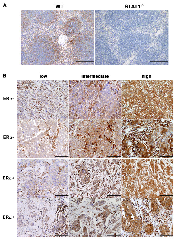
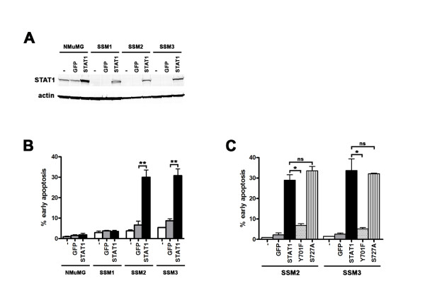
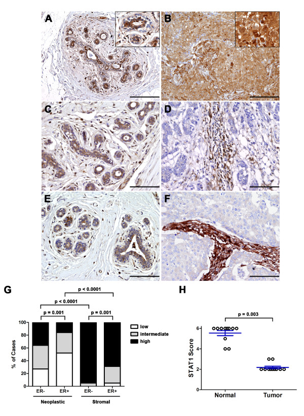
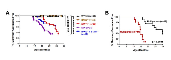
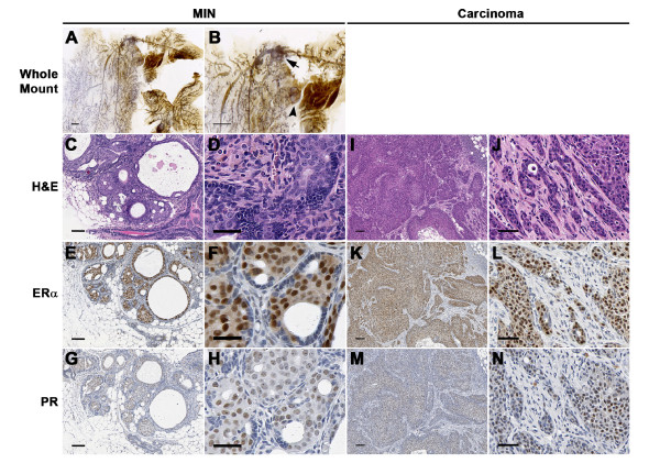
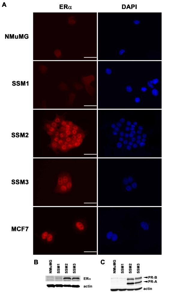
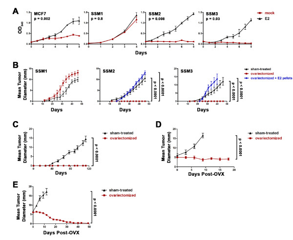
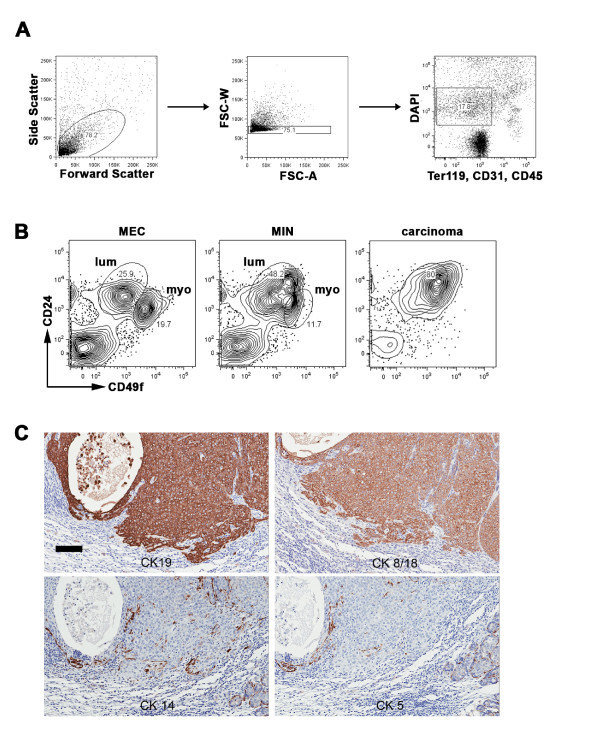
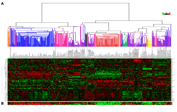
Forty-five percent (37/83) of human ERα+ and 22% (17/78) of ERα- breast cancers display undetectable or low levels of STAT1 expression in neoplastic cells. In contrast, STAT1 expression is elevated in epithelial cells of normal breast tissues adjacent to the malignant lesions, suggesting that STAT1 is selectively downregulated in the tumor cells during tumor progression. Interestingly, the expression levels of STAT1 in the tumor-infiltrating stromal cells remain elevated, indicating that single-cell resolution analysis of STAT1 level in primary breast cancer biopsies is necessary for accurate assessment. Female mice lacking functional STAT1 spontaneously develop mammary adenocarcinomas that comprise > 90% ERα+/PR+ tumor cells, and depend on estrogen for tumor engraftment and progression. Phenotypic marker analyses demonstrate that STAT1-/- mammary tumors arise from luminal epithelial cells, but not myoepithelial cells. In addition, the molecular signature of the STAT1-/- mammary tumors overlaps closely to that of human luminal breast cancers. Finally, introduction of wildtype STAT1, but not a STAT1 mutant lacking the critical Tyr701 residue, into STAT1-/- mammary tumor cells results in apoptosis, demonstrating that the tumor suppressor function of STAT1 is cell-autonomous and requires its transcriptional activity.
Our findings demonstrate that STAT1 suppresses mammary tumor formation and its expression is frequently lost during breast cancer progression. Spontaneous mammary tumors that develop in STAT1-/- mice closely recapitulate the progression, ovarian hormone responsiveness, and molecular characteristics of human luminal breast cancer, the most common subtype of human breast neoplasms, and thus represent a valuable platform for testing novel treatments and detection modalities.
尽管表达雌激素受体-α(ERα)和孕激素受体(PR)的乳腺癌是人类最常见的乳腺恶性肿瘤形式,但很难开发出具有类似甾体激素反应性的合适的小鼠模型。STAT 转录因子在乳腺肿瘤发生中发挥关键作用,但 STAT1 的精确作用尚不清楚。本文表明,一部分人类乳腺癌表现出 STAT1 表达降低,而缺乏 STAT1 的小鼠出人意料地发展出 ERα+/PR+ 乳腺肿瘤。
我们采用了多种方法,包括组织学检查、基因靶向小鼠、基因表达分析、肿瘤移植和免疫表型分析,来进行这项研究。
45%(37/83)的人类 ERα+和 22%(17/78)的 ERα-乳腺癌中,肿瘤细胞中 STAT1 的表达检测不到或很低。相比之下,STAT1 在恶性病变相邻的正常乳腺组织的上皮细胞中表达升高,表明 STAT1 在肿瘤进展过程中在肿瘤细胞中被选择性地下调。有趣的是,肿瘤浸润性基质细胞中的 STAT1 表达水平仍然升高,这表明对原发性乳腺癌活检中 STAT1 水平进行单细胞分辨率分析对于准确评估是必要的。缺乏功能性 STAT1 的雌性小鼠自发发展为乳腺腺癌,其中>90%的肿瘤细胞为 ERα+/PR+,并且依赖于雌激素进行肿瘤植入和进展。表型标志物分析表明,STAT1-/-乳腺肿瘤起源于腔上皮细胞,而不是肌上皮细胞。此外,STAT1-/-乳腺肿瘤的分子特征与人类腔型乳腺癌非常相似。最后,野生型 STAT1 的引入,但不是缺乏关键 Tyr701 残基的 STAT1 突变体的引入,导致 STAT1-/-乳腺肿瘤细胞凋亡,表明 STAT1 的肿瘤抑制功能是细胞自主的,并且需要其转录活性。
我们的研究结果表明,STAT1 抑制乳腺肿瘤的形成,并且其表达在乳腺癌进展过程中经常丢失。STAT1-/-小鼠中自发发展的乳腺肿瘤很好地重现了人类腔型乳腺癌的进展、卵巢激素反应性和分子特征,腔型乳腺癌是人类乳腺肿瘤最常见的亚型,因此代表了测试新疗法和检测方法的有价值的平台。