Zheng Linfeng, Zhang Zhuoli, Khazaie Khashayarsha, Saha Saurabh, Lewandowski Robert J, Zhang Guixiang, Larson Andrew C
Department of Radiology, First People's Hospital, Shanghai Jiaotong University, Shanghai, China; Department of Radiology, Northwestern University Feinberg School of Medicine, Chicago, Illinois, United States of America.
Department of Radiology, Northwestern University Feinberg School of Medicine, Chicago, Illinois, United States of America; Robert H. Lurie Comprehensive Cancer Center, Chicago, Illinois, United States of America.
PLoS One. 2014 Dec 30;9(12):e116204. doi: 10.1371/journal.pone.0116204. eCollection 2014.
To validate the feasibility of labeling Clostridium novyi-NT (C.novyi-NT) anaerobes with iron-oxide nanoparticles for magnetic resonance imaging (MRI) and demonstrate the potential to use MRI to visualize intra-tumoral delivery of these iron-oxide labeled C.novyi-NT during percutaneous injection procedures.
All studies were approved by IACUC. C.novyi-NT were labeled with hybrid iron-oxide Texas red nanoparticles. Growth of labeled and control samples were evaluated with optical density. Labeling was confirmed with confocal fluorescence and transmission electron microscopy (TEM). MRI were performed using a 7 Tesla scanner with T2*-weighted (T2*W) sequence. Contrast-to-noise ratio (CNR) measurements were performed for phantoms and signal-to-noise ratio (SNR) measurements performed in C57BL/6 mice (n = 12) with Panc02 xenografts before and after percutaneous injection of iron-oxide labeled C.novyi-NT. MRI was repeated 3 and 7 days post-injection. Hematoxylin-eosin (HE), Prussian blue and Gram staining of tumor specimens were performed for confirmation of intra-tumoral delivery.
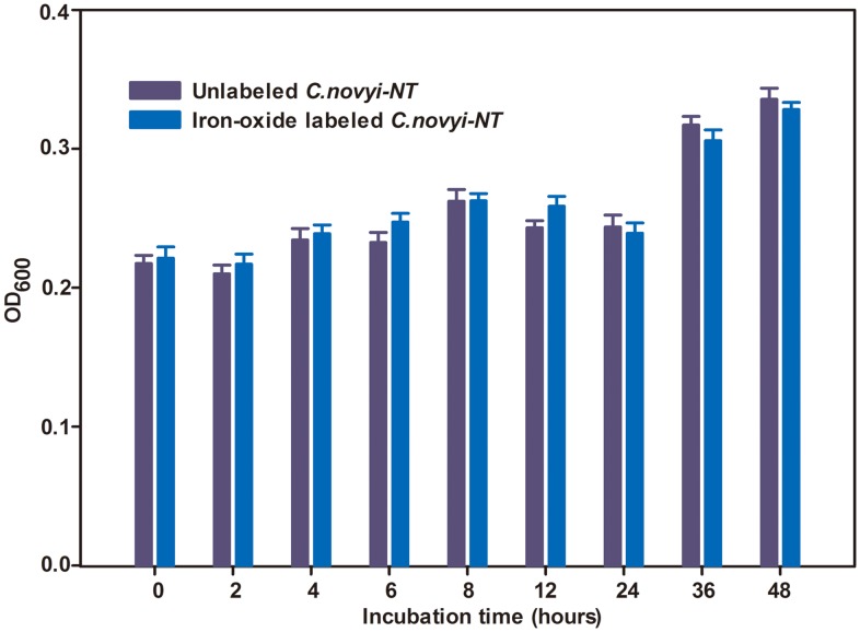
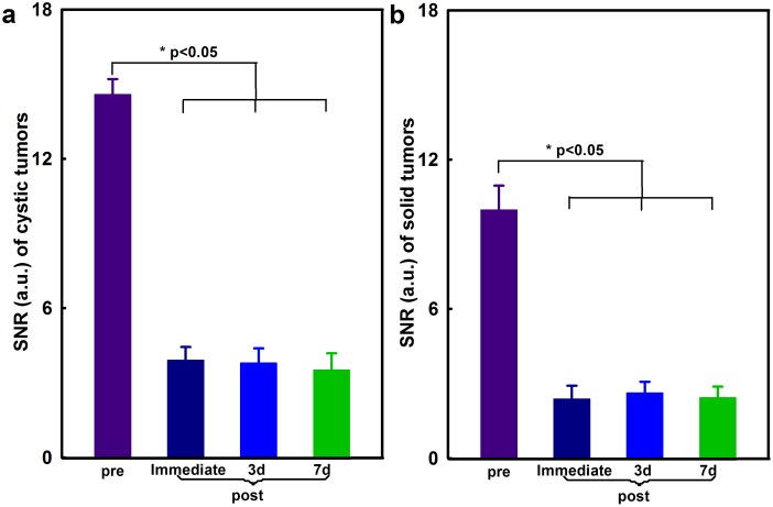
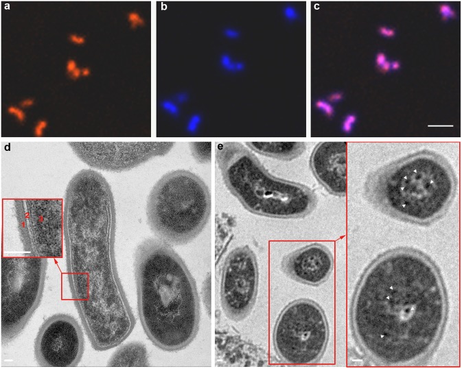
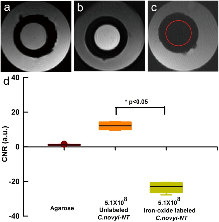
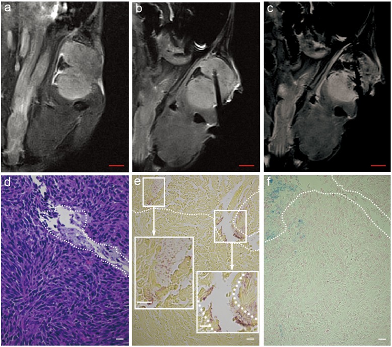
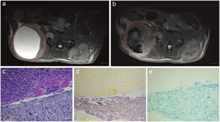
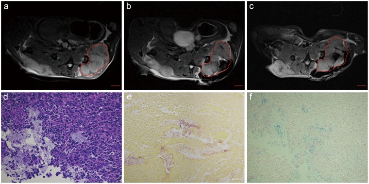
Iron-oxide labeling had no influence upon C.novyi-NT growth. The signal intensity (SI) within T2W images was significantly decreased for iron-oxide labeled C.novyi-NT phantoms compared to unlabeled controls. Under confocal fluorescence microscopy, the iron-oxide labeled C.novyi-NT exhibited a uniform red fluorescence consistent with observed regions of DAPI staining and overall labeling efficiency was 100% (all DAPI stained C.novyi-NT exhibited red fluorescence). Within TEM images, a large number iron granules were observed within the iron-oxide labeled C.novyi-NT; these were not observed within unlabeled controls. Intra-procedural MRI measurements permitted in vivo visualization of the intra-tumoral distribution of iron-oxide labeled C.novyi-NT following percutaneous injection (depicted as punctate regions of SI reductions within T2-weighted images); tumor SNR decreased significantly following intra-tumoral injection of C.novyi-NT (p<0.05); these SNR reductions were maintained at 3 and 7 day follow-up intervals. Prussian blue and Gram staining confirmed presence of the iron-oxide labeled anaerobes.
C.novyi-NT can be labeled with iron-oxide nanoparticles for MRI visualization of intra-tumoral deposition following percutaneous injection during bacteriolytic therapy.
验证用氧化铁纳米颗粒标记诺维氏梭菌 NT(C.novyi-NT)厌氧菌用于磁共振成像(MRI)的可行性,并证明在经皮注射过程中利用 MRI 可视化这些氧化铁标记的 C.novyi-NT 在肿瘤内递送的潜力。
所有研究均经机构动物护理与使用委员会(IACUC)批准。C.novyi-NT 用混合氧化铁德克萨斯红纳米颗粒进行标记。通过光密度评估标记样本和对照样本的生长情况。用共聚焦荧光显微镜和透射电子显微镜(TEM)确认标记情况。使用配备 T2加权(T2W)序列的 7 特斯拉扫描仪进行 MRI 检查。对体模进行对比噪声比(CNR)测量,对患有 Panc02 异种移植瘤的 C57BL/6 小鼠(n = 12)在经皮注射氧化铁标记的 C.novyi-NT 前后进行信噪比(SNR)测量。在注射后 3 天和 7 天重复进行 MRI 检查。对肿瘤标本进行苏木精 - 伊红(HE)、普鲁士蓝和革兰氏染色以确认肿瘤内递送情况。
氧化铁标记对 C.novyi-NT 的生长没有影响。与未标记的对照相比,氧化铁标记的 C.novyi-NT 体模在 T2W 图像中的信号强度(SI)显著降低。在共聚焦荧光显微镜下,氧化铁标记的 C.novyi-NT 呈现均匀的红色荧光,与观察到的 DAPI 染色区域一致,总体标记效率为 100%(所有 DAPI 染色的 C.novyi-NT 均呈现红色荧光)。在 TEM 图像中,在氧化铁标记的 C.novyi-NT 内观察到大量铁颗粒;在未标记的对照中未观察到这些颗粒。术中 MRI 测量允许在经皮注射后对氧化铁标记的 C.novyi-NT 在肿瘤内的分布进行体内可视化(在 T2加权图像中表现为 SI 降低的点状区域);肿瘤内注射 C.novyi-NT 后肿瘤 SNR 显著降低(p<0.05);这些 SNR 降低在 3 天和 7 天的随访间隔中持续存在。普鲁士蓝和革兰氏染色证实了氧化铁标记厌氧菌的存在。
C.novyi-NT 可用氧化铁纳米颗粒进行标记,以便在溶菌治疗期间经皮注射后通过 MRI 可视化肿瘤内沉积情况。