Harston George W J, Tee Yee Kai, Blockley Nicholas, Okell Thomas W, Thandeswaran Sivarajan, Shaya Gabriel, Sheerin Fintan, Cellerini Martino, Payne Stephen, Jezzard Peter, Chappell Michael, Kennedy James
1 Acute Stroke Programme, Radcliffe Department of Medicine, University of Oxford, UK.
2 Institute of Biomedical Engineering, Department of Engineering Science, University of Oxford, UK 3 Department of Mechatronics and Biomedical Engineering, Lee Kong Chian Faculty of Engineering and Science, Universiti Tunku Abdul Rahman, Malaysia.
Brain. 2015 Jan;138(Pt 1):36-42. doi: 10.1093/brain/awu374.
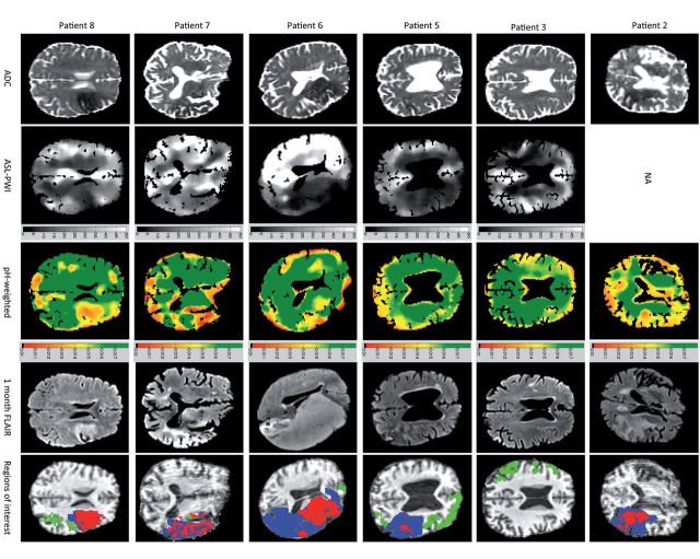
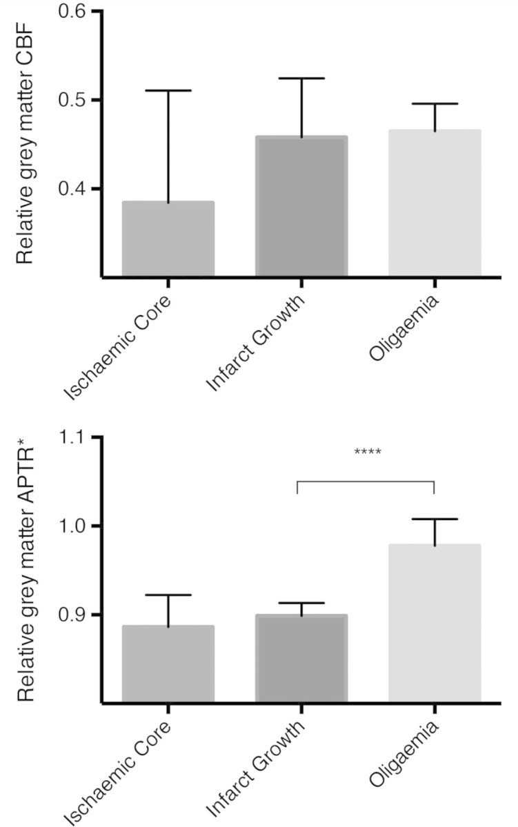
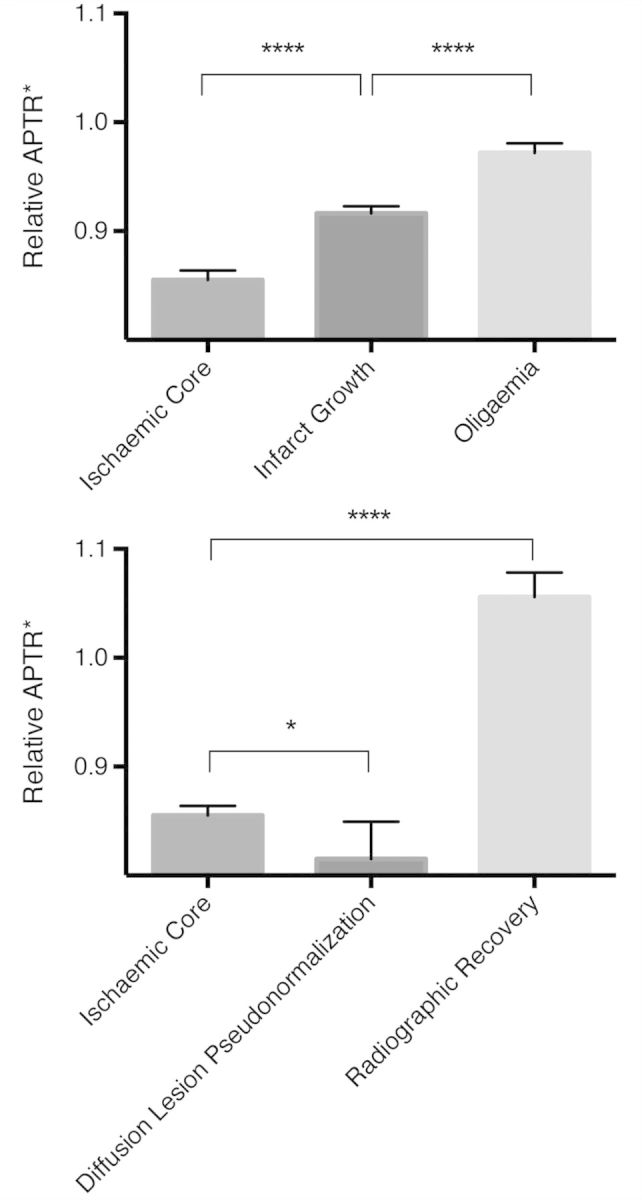
The original concept of the ischaemic penumbra suggested imaging of regional cerebral blood flow and metabolism would be required to identify tissue that may benefit from intervention. Amide proton transfer magnetic resonance imaging, a chemical exchange saturation transfer technique, has been used to derive cerebral intracellular pH in preclinical stroke models and has been proposed as a metabolic marker of ischaemic penumbra. In this proof of principle clinical study, we explored the potential of this pH-weighted magnetic resonance imaging technique at tissue-level. Detailed voxel-wise analysis was performed on data from a prospective cohort of 12 patients with acute ischaemic stroke. Voxels within ischaemic core had a more severe intracellular acidosis than hypoperfused tissue recruited to the final infarct (P < 0.0001), which in turn was more acidotic than hypoperfused tissue that survived (P < 0.0001). In addition, when confined to the grey matter perfusion deficit, intracellular pH (P < 0.0001), but not cerebral blood flow (P = 0.31), differed between tissue that infarcted and tissue that survived. Within the presenting apparent diffusion coefficient lesion, intracellular pH differed between tissue with early apparent diffusion lesion pseudonormalization and tissue with true radiographic recovery. These findings support the need for further investigation of pH-weighted imaging in patients with acute ischaemic stroke.
缺血半暗带的最初概念表明,需要对局部脑血流和代谢进行成像,以识别可能从干预中获益的组织。酰胺质子转移磁共振成像作为一种化学交换饱和转移技术,已被用于在临床前卒中模型中测定脑细胞内pH值,并被提议作为缺血半暗带的代谢标志物。在这项原理验证临床研究中,我们在组织水平上探索了这种pH加权磁共振成像技术的潜力。对12例急性缺血性卒中患者的前瞻性队列数据进行了详细的体素分析。缺血核心内的体素比最终梗死灶中募集的灌注不足组织有更严重的细胞内酸中毒(P<0.0001),而后者又比存活的灌注不足组织酸性更强(P<0.0001)。此外,当局限于灰质灌注缺损时,梗死组织和存活组织之间的细胞内pH值存在差异(P<0.0001),但脑血流量无差异(P=0.31)。在呈现的表观扩散系数病变范围内,早期表观扩散病变假性正常化的组织与真正影像学恢复的组织之间的细胞内pH值存在差异。这些发现支持对急性缺血性卒中患者进行pH加权成像的进一步研究。