Grote Anne, Abbas Mahmoud, Linder Nina, Kreipe Hans H, Lundin Johan, Feuerhake Friedrich
Diagn Pathol. 2014;9 Suppl 1(Suppl 1):S11. doi: 10.1186/1746-1596-9-S1-S11. Epub 2014 Dec 19.
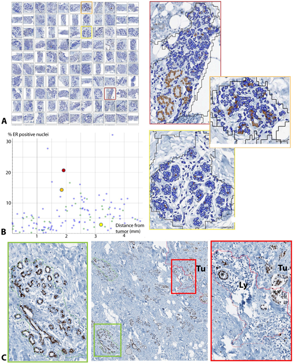
Comprehensive spatial assessment of hormone receptor immunohistochemistry staining in digital whole slide images of breast cancer requires accurate detection of positive nuclei within biologically relevant regions of interest. Herein, we propose a combination of automated region labeling at low resolution and subsequent detailed tissue evaluation of subcellular structures in lobular structures adjacent to breast cancer, as a proof of concept for the approach to analyze estrogen and progesterone receptor expression in the spatial context of surrounding tissue.
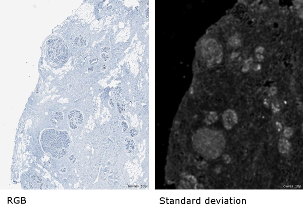
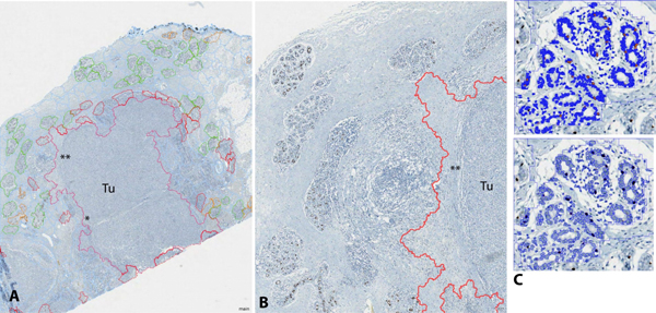
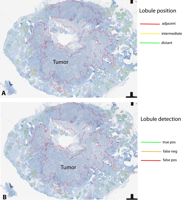
Routinely processed paraffin sections of hormone receptor-negative ductal invasive breast cancer were stained for estrogen and progesterone receptor by immunohistochemistry. Digital whole slides were analyzed using commercially available image analysis software for advanced object-based analysis, applying textural, relational, and geometrical features. Mammary gland lobules were targeted as regions of interest for analysis at subcellular level in relation to their distance from coherent tumor as neighboring relevant tissue compartment. Lobule detection quality was evaluated visually by a pathologist.
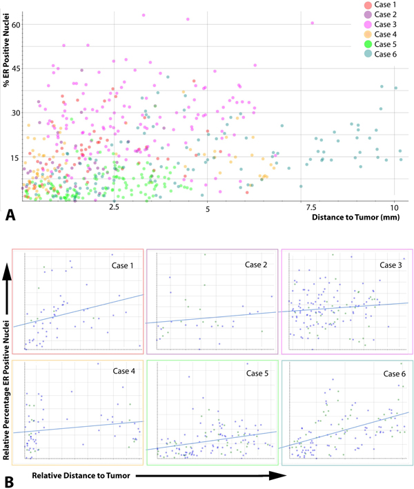
After rule set optimization in an estrogen receptor-stained training set, independent test sets (progesterone and estrogen receptor) showed acceptable detection quality in 33% of cases. Presence of disrupted lobular structures, either by brisk inflammatory infiltrate, or diffuse tumor infiltration, was common in cases with lower detection accuracy. Hormone receptor detection tended towards higher percentage of positively stained nuclei in lobules distant from the tumor border as compared to areas adjacent to the tumor. After adaptations of image analysis, corresponding evaluations were also feasible in hormone receptor positive breast cancer, with some limitations of automated separation of mammary epithelial cells from hormone receptor-positive tumor cells.
As a proof of concept for object-oriented detection of steroid hormone receptors in their spatial context, we show that lobular structures can be classified based on texture-based image features, unless brisk inflammatory infiltration disrupts the normal morphological structure of the tubular gland epithelium. We consider this approach as prototypic for detection and spatial analysis of nuclear markers in defined regions of interest. We conclude that advanced image analysis at this level of complexity requires adaptation to the individual tumor phenotypes and morphological characteristics of the tumor environment.
在乳腺癌数字全切片图像中对激素受体免疫组化染色进行全面的空间评估,需要在生物学相关的感兴趣区域内准确检测阳性细胞核。在此,我们提出一种结合低分辨率自动区域标记以及随后对乳腺癌相邻小叶结构中亚细胞结构进行详细组织评估的方法,作为在周围组织空间背景下分析雌激素和孕激素受体表达方法的概念验证。
对激素受体阴性的导管浸润性乳腺癌常规处理的石蜡切片进行雌激素和孕激素受体免疫组化染色。使用市售图像分析软件对数字全切片进行分析,以进行基于对象的高级分析,应用纹理、关系和几何特征。将乳腺小叶作为感兴趣区域,在亚细胞水平上分析其与连贯肿瘤的距离,将其作为相邻相关组织隔室。由病理学家对小叶检测质量进行视觉评估。
在雌激素受体染色的训练集中进行规则集优化后,独立测试集(孕激素和雌激素受体)在33%的病例中显示出可接受的检测质量。在检测准确性较低的病例中,小叶结构被活跃的炎性浸润或弥漫性肿瘤浸润破坏的情况很常见。与肿瘤相邻区域相比,远离肿瘤边界的小叶中激素受体检测的阳性染色细胞核百分比往往更高。在对图像分析进行调整后,在激素受体阳性乳腺癌中也可行相应评估,但在将乳腺上皮细胞与激素受体阳性肿瘤细胞自动分离方面存在一些局限性。
作为在空间背景下对类固醇激素受体进行面向对象检测的概念验证,我们表明,除非活跃的炎性浸润破坏管状腺上皮的正常形态结构,否则可以基于基于纹理的图像特征对小叶结构进行分类。我们认为这种方法是在定义的感兴趣区域中检测和空间分析核标记的原型。我们得出结论,在这种复杂程度下的高级图像分析需要适应个体肿瘤表型和肿瘤环境的形态特征。