Lee Ju Hee, Choung Sorim, Kim Ji Min, Lee Jung Uee, Kim Koon Soon, Kim Hyun Jin, Jeong Jae-Wook, Ku Bon Jeong
Department of Internal Medicine, Chungnam National University School of Medicine, 282 Munhwa-ro, Jung-gu, Daejeon 301-721, Republic of Korea.
Department of Pathology, Daejeon St. Mary's Hospital, College of Medicine, The Catholic University of Korea, 222 Banpo-daero, Seocho-gu, Seoul 137-701, Republic of Korea.
Dis Markers. 2014;2014:549054. doi: 10.1155/2014/549054. Epub 2014 Dec 10.
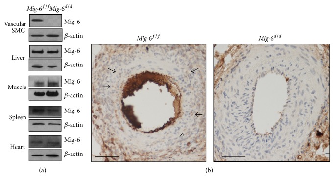
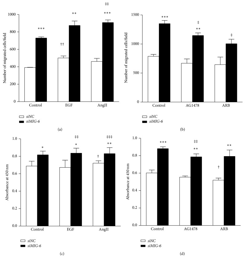
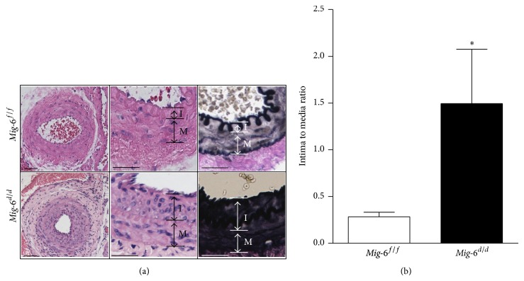
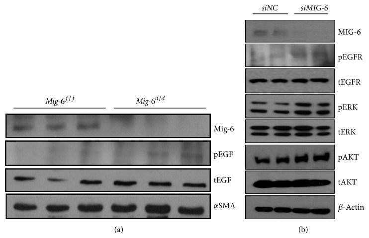
Although advances in vascular interventions can reduce the mortality associated with cardiovascular disease, neointimal hyperplasia remains a clinically significant obstacle limiting the success of current interventions. Identification of signaling pathways involved in migration and proliferation of vascular smooth muscle cells (SMCs) is an important approach for the development of modalities to combat this disease. Herein we investigate the role of an immediate early response gene, mitogen-inducible gene-6 (Mig-6), in the development of neointimal hyperplasia using vascular smooth muscle specific Mig-6 knockout mice. We induced endoluminal injury to one side of femoral artery by balloon dilatation in both Mig-6 knockout and control mice. Four weeks following injury, the artery of Mig-6 knockout mice demonstrated a 5.3-fold increase in the neointima/media ratio compared with control mice (P = 0.04). In addition, Mig-6 knockout vascular SMCs displayed an increase in both cell migration and proliferation compared with wild-type SMCs. Taken together, our data suggest that Mig-6 plays a critical role in the development of atherosclerosis. This finding provides new insight into the development of more effective ways to treat and prevent neointimal hyperplasia, particularly in-stent restenosis after percutaneous vascular intervention.
尽管血管介入技术的进步可以降低心血管疾病相关的死亡率,但新生内膜增生仍然是限制当前介入治疗成功的一个具有临床意义的障碍。识别参与血管平滑肌细胞(SMC)迁移和增殖的信号通路是开发对抗这种疾病方法的重要途径。在此,我们使用血管平滑肌特异性Mig-6基因敲除小鼠,研究即刻早期反应基因丝裂原诱导基因-6(Mig-6)在新生内膜增生发展中的作用。我们通过球囊扩张对Mig-6基因敲除小鼠和对照小鼠的一侧股动脉造成腔内损伤。损伤后四周,与对照小鼠相比,Mig-6基因敲除小鼠的动脉新生内膜/中膜比值增加了5.3倍(P = 0.04)。此外,与野生型SMC相比,Mig-6基因敲除的血管SMC在细胞迁移和增殖方面均有所增加。综上所述,我们的数据表明Mig-6在动脉粥样硬化的发展中起关键作用。这一发现为开发更有效的治疗和预防新生内膜增生的方法提供了新的见解,特别是经皮血管介入术后的支架内再狭窄。