Jonsson Joakim H, Akhtari Mohammad M, Karlsson Magnus G, Johansson Adam, Asklund Thomas, Nyholm Tufve
Department of Radiation Sciences, Umeå University, Umeå, SE-901 87, Sweden.
Radiat Oncol. 2015 Jan 10;10:13. doi: 10.1186/s13014-014-0308-1.
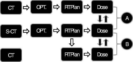
In this pilot study we evaluated the performance of a substitute CT (s-CT) image derived from MR data of the brain, as a basis for optimization of intensity modulated rotational therapy, final dose calculation and derivation of reference images for patient positioning.
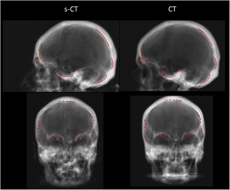
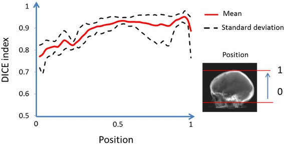
S-CT images were created using a Gaussian mixture regression model on five patients previously treated with radiotherapy. Optimizations were compared using D max, D min, D median and D mean measures for the target volume and relevant risk structures. Final dose calculations were compared using gamma index with 1%/1 mm and 3%/3 mm acceptance criteria. 3D geometric evaluation was conducted using the DICE similarity coefficient for bony structures. 2D geometric comparison of digitally reconstructed radiographs (DRRs) was performed by manual delineation of relevant structures on the s-CT DRR that were transferred to the CT DRR and compared by visual inspection.
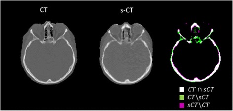
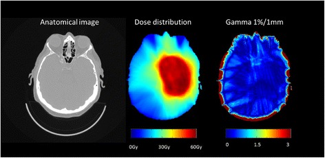
Differences for the target volumes in optimization comparisons were small in general, e.g. a mean difference in both D min and D max within ±0.3%. For the final dose calculation gamma evaluations, 100% of the voxels passed the 1%/1 mm criterion within the PTV. Within the entire external volume between 99.4% and 100% of the voxels passed the 3%/3 mm criterion. In the 3D geometric comparison, the DICE index varied between approximately 0.8-0.9, depending on the position in the skull. In the 2D DRR comparisons, no appreciable visual differences were found.
Even though the present work involves a limited number of patients, the results provide a strong indication that optimization and dose calculation based on s-CT data is accurate regarding both geometry and dosimetry.
在这项初步研究中,我们评估了从脑部磁共振(MR)数据得出的替代CT(s-CT)图像的性能,作为强度调制旋转治疗优化、最终剂量计算以及患者定位参考图像推导的基础。
使用高斯混合回归模型为五名先前接受过放射治疗的患者创建s-CT图像。使用靶区体积和相关危险结构的D max、D min、D median和D mean测量值比较优化结果。使用1%/1 mm和3%/3 mm接受标准的伽马指数比较最终剂量计算结果。使用骨结构的DICE相似系数进行三维几何评估。通过在s-CT数字重建X线片(DRR)上手动描绘相关结构并将其转移到CT DRR上,通过目视检查进行二维几何比较。
优化比较中靶区体积的差异总体较小,例如D min和D max的平均差异在±0.3%以内。对于最终剂量计算的伽马评估,100%的体素在计划靶体积(PTV)内通过1%/1 mm标准。在整个外部体积中,99.4%至100%的体素通过3%/3 mm标准。在三维几何比较中,DICE指数根据在颅骨中的位置在约0.8 - 0.9之间变化。在二维DRR比较中,未发现明显的视觉差异。
尽管目前的工作涉及的患者数量有限,但结果有力地表明基于s-CT数据的优化和剂量计算在几何形状和剂量学方面都是准确的。