Orsini G, Majorana A, Mazzoni A, Putignano A, Falconi M, Polimeni A, Breschi L
Polytechnic University of Marche.
Eur J Histochem. 2014 Dec 1;58(4):2405. doi: 10.4081/ejh.2014.2405.
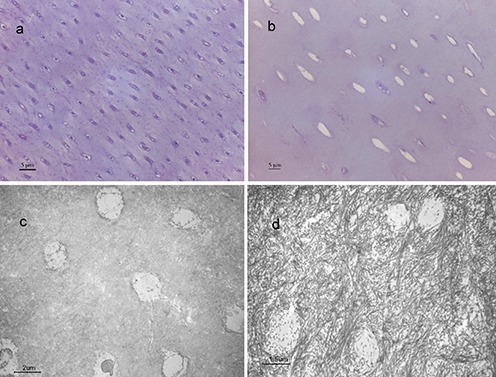
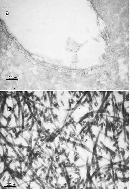
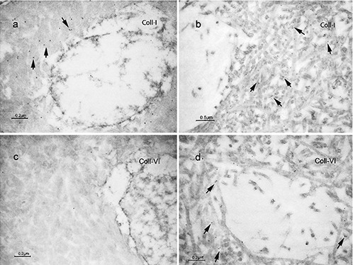
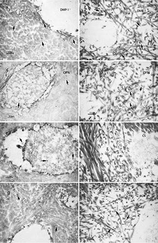
Dentinogenesis imperfecta determines structural alterations of the collagen structure still not completely elucidated. Immunohistochemical analysis was used to assay Type I and VI collagen, various non-collagenous proteins distribution in human primary teeth from healthy patients or from patients affected by type I dentinogenesis imperfecta (DGI-I) associated with osteogenesis imperfecta (OI). In sound primary teeth, an organized well-known ordered pattern of the type I collagen fibrils was found, whereas atypical and disorganized fibrillar structures were observed in dentin of DGI-I affected patients. Expression of type I collagen was observed in both normal and affected primary teeth, although normal dentin stained more uniformly than DGI-I affected dentin. Reactivity of type VI collagen was significantly lower in normal teeth than in dentin from DGI-I affected patients (P<0.05). Expressions of dentin matrix protein (DMP)-1 and osteopontin (OPN) were observed in both normal dentin and dentin from DGI-I affected patients, without significant differences, being DMP1 generally more abundantly expressed. Immunolabeling for chondroitin sulfate (CS) and biglycan (BGN) was weaker in dentin from DGI-I-affected patients compared to normal dentin, this decrease being significant only for CS. This study shows ultrastructural alterations in dentin obtained from patients affected by DGI-I, supported by immunocytochemical assays of different collagenous and non-collagenous proteins.
牙本质发育不全决定了胶原结构的结构改变,但其仍未完全阐明。免疫组织化学分析用于检测健康患者或患有与成骨不全(OI)相关的I型牙本质发育不全(DGI-I)患者的人乳牙中I型和VI型胶原、各种非胶原蛋白质的分布。在健康乳牙中,发现了I型胶原纤维的一种有组织的、众所周知的有序模式,而在DGI-I患者的牙本质中观察到了非典型和无序的纤维结构。在正常和患病乳牙中均观察到I型胶原的表达,尽管正常牙本质的染色比DGI-I患者的牙本质更均匀。正常牙齿中VI型胶原的反应性明显低于DGI-I患者牙本质中的反应性(P<0.05)。在正常牙本质和DGI-I患者牙本质中均观察到牙本质基质蛋白(DMP)-1和骨桥蛋白(OPN)的表达,无显著差异,DMP1通常表达更丰富。与正常牙本质相比,DGI-I患者牙本质中硫酸软骨素(CS)和双糖链蛋白聚糖(BGN)的免疫标记较弱,这种降低仅对CS有显著意义。本研究显示了DGI-I患者牙本质的超微结构改变,并得到了不同胶原和非胶原蛋白质免疫细胞化学分析的支持。