Sacharz Joanna, Bryan Samantha J, Yu Jianfeng, Burroughs Nigel J, Spence Edward M, Nixon Peter J, Mullineaux Conrad W
School of Biological and Chemical Sciences, Queen Mary University of London, Mile End Road, London, E1 4NS, UK.
Mol Microbiol. 2015 May;96(3):448-62. doi: 10.1111/mmi.12940. Epub 2015 Feb 11.
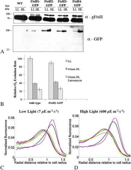
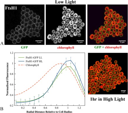
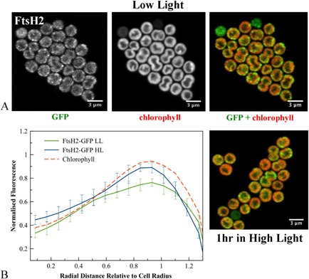
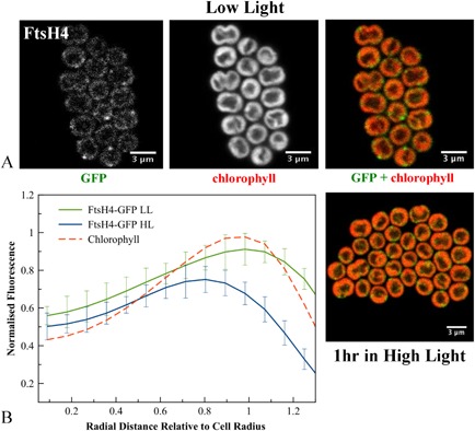
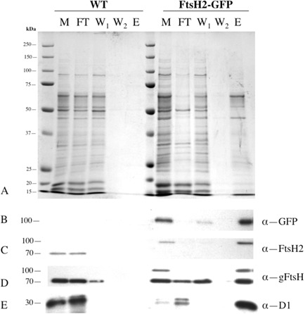
In cyanobacteria and chloroplasts, exposure to HL damages the photosynthetic apparatus, especially the D1 subunit of Photosystem II. To avoid chronic photoinhibition, a PSII repair cycle operates to replace damaged PSII subunits with newly synthesised versions. To determine the sub-cellular location of this process, we examined the localisation of FtsH metalloproteases, some of which are directly involved in degrading damaged D1. We generated transformants of the cyanobacterium Synechocystis sp. PCC6803 expressing GFP-tagged versions of its four FtsH proteases. The ftsH2-gfp strain was functional for PSII repair under our conditions. Confocal microscopy shows that FtsH1 is mainly in the cytoplasmic membrane, while the remaining FtsH proteins are in patches either in the thylakoid or at the interface between the thylakoid and cytoplasmic membranes. HL exposure which increases the activity of the Photosystem II repair cycle led to no detectable changes in FtsH distribution, with the FtsH2 protease involved in D1 degradation retaining its patchy distribution in the thylakoid membrane. We discuss the possibility that the FtsH2-GFP patches represent Photosystem II 'repair zones' within the thylakoid membranes, and the possible advantages of such functionally specialised membrane zones. Anti-GFP affinity pull-downs provide the first indication of the composition of the putative repair zones.
在蓝细菌和叶绿体中,暴露于高光(HL)会损害光合机构,尤其是光系统II的D1亚基。为避免慢性光抑制,光系统II修复循环发挥作用,用新合成的亚基替换受损的光系统II亚基。为确定该过程的亚细胞定位,我们研究了FtsH金属蛋白酶的定位,其中一些直接参与降解受损的D1。我们构建了集胞藻属蓝细菌PCC6803的转化体,其表达四种FtsH蛋白酶的绿色荧光蛋白(GFP)标记版本。在我们的条件下,ftsH2 - gfp菌株对光系统II修复具有功能。共聚焦显微镜显示,FtsH1主要位于细胞质膜中,而其余的FtsH蛋白则以斑块形式存在于类囊体中或类囊体与细胞质膜的界面处。高光暴露增加了光系统II修复循环的活性,但FtsH分布未检测到变化,参与D1降解的FtsH2蛋白酶在类囊体膜中仍保持其斑块状分布。我们讨论了FtsH2 - GFP斑块代表类囊体膜内光系统II“修复区”的可能性,以及这种功能特化膜区的潜在优势。抗GFP亲和下拉实验首次揭示了假定修复区的组成情况。