Zhou Hui, Luo Mei, Wen Yige, Ma Andi, Luo Yongzhong, Yi Qing, Chen Jianhua, Xiao Ling
Hunan Cancer Hospital, the Affiliated Cancer Hospital of Xiangya School of Medicine, Central South University, Changsha 410013, China;State Key Laboratory of Medical Genetics Central South University, Changsha 410078, China.
Hunan Cancer Hospital, the Affiliated Cancer Hospital of Xiangya School of Medicine, Central South University, Changsha 410013, China.
Zhongguo Fei Ai Za Zhi. 2015 Jan;18(1):8-15. doi: 10.3779/j.issn.1009-3419.2015.01.02.
It has been proven that toll-like receptor 5 (TLR5) plaied an important role in the development of tumor. In our previous study, we found that the expression of TLR5 was remarkably higher in non-small cell lung cancer (NSCLC) tissues than that in normal tissues, but the activation of TLR5 signaling pathway in NSCLC was still unknown. The aim of this study is to investigate the expression of TLR5 in different types of NSCLC cell lines, and analyze the activity of the signaling pathway after stimulated by its specific exogenous ligand flagellin.
The TLR5 protein was detected by immunofluorescence and Western blot in three kinds of NSCLC cell lines, and the TLR5 mRNA was detected by RT-PCR. Select the cell line of TLR5 highest expression as the research object, and select the suitable concentration of flagellin. NF-κB luciferase activity was detected to validate the TLR5 activation pathway through inhibitory signaling pathways by 0 μg/mL, 0.01 μg/mL, 0.1 μg/mL, 1 μg/mL, 10 μg/mL TLR5 antibody. The chosen cell line was transfected by TLR5 shRNA plasmid, and p-IKBα, IKBα, p-ERK1/2, ERK1/2 and p-JNK of untrasfected and transfected cells were detected in the activity of TLR5 signaling pathway by Western blot at 0 min, 10 min, 30 min and 60 min, respectively.
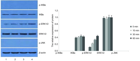
The expression of TLR5 was the highest in the lung adenocarcinoma cell line SPC-A-1 by immunofluorescence, mainly expressed on the cell membrane. NF-κB luciferase activity of SPC-A-1 cells was the highest, and the activity was increased in a dose-dependent manner. 0.1 μg/mL flagellin could significantly increase the NF-κB luciferase activity (P<0.05), while its activity could be inhibited by the TLR5 antibody in a negative correlation. Treated by 0.1 μg/mL flagellin, compared with that of 0 min group, the levels of p-IKBα, p-ERK1/2, p-JNK of SPC-A-1 cells increased significantly after 10 min, reached the peak at 30 min, and declined at 60 min (P<0.05). Compared with that of 10 min and 60 min group, the levels of p-IKBα, p-ERK1/2, p-JNK significantly increased at 30 min (P<0.05). While the levels of IKBα, ERK1/2 at 0 min, 10 min, 30 min and 60 min had no significant changes (P>0.05). SPC-A-1 cells transfected TLR5-shRNA were also stimulated by flagellin (0.1 μg/mL). At 0 min, 10 min, 30 min and 60 min, p-IKBα and p-JNK proteins could not be detected, and the levels of IKBα and ERK1/2 had no significant changes (P>0.05), but the levels of p-ERK1/2 significantly increased as time went on (P<0.05).
Exogenous ligand flagellin can activate TLR5 protein in NSCLC cell lines and initiate downstream signaling pathways. It may be relative to the development of NSCLC.
已证实Toll样受体5(TLR5)在肿瘤发生发展中起重要作用。在我们之前的研究中,发现非小细胞肺癌(NSCLC)组织中TLR5的表达明显高于正常组织,但NSCLC中TLR5信号通路的激活情况仍不清楚。本研究旨在探讨TLR5在不同类型NSCLC细胞系中的表达,并分析其特异性外源性配体鞭毛蛋白刺激后信号通路的活性。
采用免疫荧光和蛋白质印迹法检测三种NSCLC细胞系中TLR5蛋白,采用逆转录-聚合酶链反应(RT-PCR)检测TLR5 mRNA。选择TLR5表达最高的细胞系作为研究对象,选择合适浓度的鞭毛蛋白。通过0 μg/mL、0.01 μg/mL、0.1 μg/mL、1 μg/mL、10 μg/mL TLR5抗体抑制信号通路,检测NF-κB荧光素酶活性以验证TLR5激活途径。用TLR5 shRNA质粒转染所选细胞系,分别在0 min、10 min、30 min和60 min通过蛋白质印迹法检测未转染和转染细胞在TLR5信号通路活性中的p-IKBα、IKBα、p-ERK1/2、ERK1/2和p-JNK。
免疫荧光显示肺腺癌细胞系SPC-A-1中TLR5表达最高,主要表达于细胞膜。SPC-A-1细胞的NF-κB荧光素酶活性最高,且活性呈剂量依赖性增加。0.1 μg/mL鞭毛蛋白可显著增加NF-κB荧光素酶活性(P<0.05),而其活性可被TLR5抗体呈负相关抑制。用0.1 μg/mL鞭毛蛋白处理后,与0 min组相比,SPC-A-1细胞的p-IKBα、p-ERK1/2、p-JNK水平在10 min后显著升高,30 min达到峰值,60 min下降(P<0.05)。与10 min和60 min组相比,30 min时p-IKBα、p-ERK1/2、p-JNK水平显著升高(P<0.05)。而0 min、10 min、30 min和60 min时IKBα、ERK1/2水平无显著变化(P>0.05)。转染TLR5-shRNA的SPC-A-1细胞也用鞭毛蛋白(0.1 μg/mL)刺激。在0 min、10 min、30 min和60 min时,未检测到p-IKBα和p-JNK蛋白,IKBα和ERK1/2水平无显著变化(P>0.05),但p-ERK1/2水平随时间推移显著升高(P<0.05)。
外源性配体鞭毛蛋白可激活NSCLC细胞系中的TLR5蛋白并启动下游信号通路。这可能与NSCLC的发生发展有关。