Baghaban Eslaminejad Mohamadreza, Bordbar Sima
Department of Stem Cells and Developmental Biology, Cell Science Research Center, Royan Institute for Stem Cell Biology and Technology, Academic Center for Education, Culture and Research (ACECR), Tehran, Iran.
Vet Res Forum. 2012 Summer;3(3):159-65.
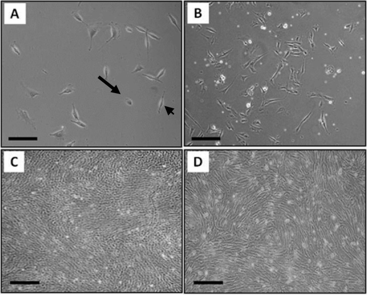
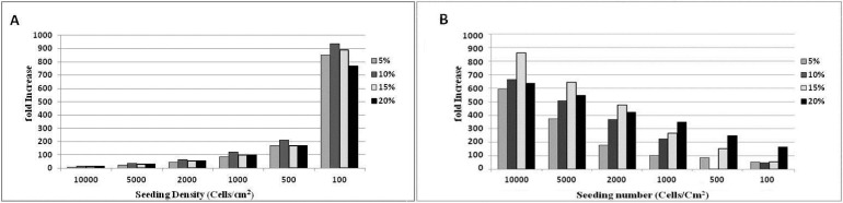
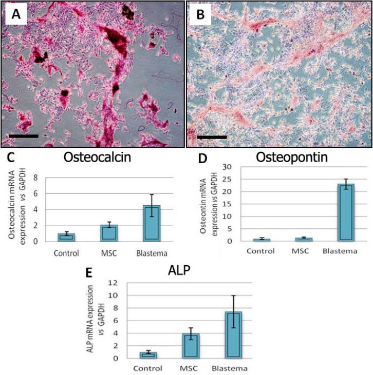
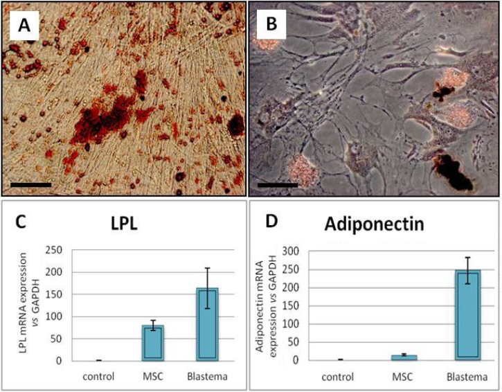
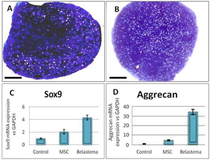
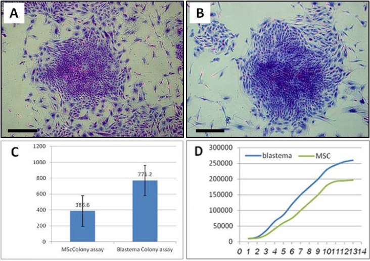
Rabbits have the capacity to regenerate holes in their ears by forming a blastema, a tissue that is made up of a group of undifferentiated cells. The purpose of the present study was to isolate and characterize blastema progenitor cells and compare them with marrow mesenchymal stem cells (MSCs). Five New Zealand white male rabbits were used in the present study. A 2-mm hole was created in the animal ears. After 4 days, the blastema ring formed in the periphery of the hole was removed and cultivated. The cells were expanded through several subcultures and compared with the MSCs derived from the marrow of same animal in terms of in vitro differentiation capacity, growth kinetics and culture requirements for optimal proliferation. The primary cultures from both cells tended to be heterogeneous. Fibroblastic cells became progressively dominant with advancing passages. Similar to MSCs blastema passaged-3 cells succeeded to differentiate into bone, cartilage and adipose cell lineages. Even lineage specific genes tended to express in higher level in blastema cells compared to MSCs (p < 0.05). Moreover blastema cells appeared more proliferative; producing more colonies (p < 0.05). While blastema cells showed extensive proliferation in 15% fetal bovine serum (FBS), MSCs displayed higher expansion rate at 10% FBS. In conclusion, blastema from rabbit ear contains a population of fibroblastic cells much similar in characteristic to bone marrow mesenchymal stem cells. However, the two cells were different in the level of lineage-specific gene expression, the growth curve characteristics and the culture requirements.
兔子能够通过形成芽基来再生耳朵上的孔洞,芽基是一种由一群未分化细胞组成的组织。本研究的目的是分离和鉴定芽基祖细胞,并将它们与骨髓间充质干细胞(MSCs)进行比较。本研究使用了5只新西兰雄性白兔。在动物耳朵上制造一个2毫米的孔洞。4天后,将在孔洞周边形成的芽基环移除并进行培养。通过多次传代培养使细胞扩增,并在体外分化能力、生长动力学和最佳增殖所需的培养条件方面,将这些细胞与来自同一动物骨髓的MSCs进行比较。两种细胞的原代培养物都倾向于具有异质性。随着传代次数的增加,成纤维细胞逐渐占主导地位。与MSCs相似,传代3次的芽基细胞成功分化为骨、软骨和脂肪细胞谱系。甚至与MSCs相比,谱系特异性基因在芽基细胞中的表达水平往往更高(p<0.05)。此外,芽基细胞表现出更强的增殖能力;产生更多的集落(p<0.05)。当芽基细胞在15%胎牛血清(FBS)中表现出广泛增殖时,MSCs在10%FBS中显示出更高的扩增率。总之,兔耳芽基含有一群成纤维细胞,其特征与骨髓间充质干细胞非常相似。然而,这两种细胞在谱系特异性基因表达水平、生长曲线特征和培养条件方面存在差异。