Ma Yizhou, Koyama Maki S, Milham Michael P, Castellanos F Xavier, Quinn Brian T, Pardoe Heath, Wang Xiuyuan, Kuzniecky Ruben, Devinsky Orrin, Thesen Thomas, Blackmon Karen
Department of Neurology, Comprehensive Epilepsy Center, School of Medicine, New York University, New York, NY, USA ; Department of Psychology, New York University, New York, NY, USA.
Child Mind Institute, New York, NY, USA ; Nathan Kline Institute for Psychiatric Research, Orangeburg, NY, USA.
Neuroimage Clin. 2014 Nov 18;7:177-86. doi: 10.1016/j.nicl.2014.11.005. eCollection 2015.
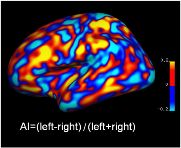
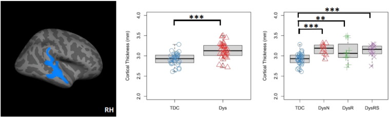
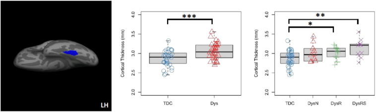
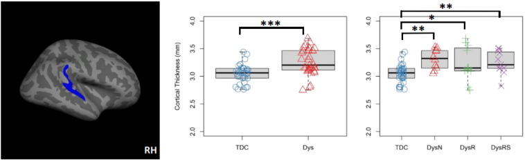
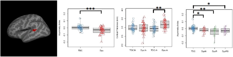
Abnormalities in cortical structure are commonly observed in children with dyslexia in key regions of the "reading network." Whether alteration in cortical features reflects pathology inherent to dyslexia or environmental influence (e.g., impoverished reading experience) remains unclear. To address this question, we compared MRI-derived metrics of cortical thickness (CT), surface area (SA), gray matter volume (GMV), and their lateralization across three different groups of children with a historical diagnosis of dyslexia, who varied in current reading level. We compared three dyslexia subgroups with: (1) persistent reading and spelling impairment; (2) remediated reading impairment (normal reading scores), and (3) remediated reading and spelling impairments (normal reading and spelling scores); and a control group of (4) typically developing children. All groups were matched for age, gender, handedness, and IQ. We hypothesized that the dyslexia group would show cortical abnormalities in regions of the reading network relative to controls, irrespective of remediation status. Such a finding would support that cortical abnormalities are inherent to dyslexia and are not a consequence of abnormal reading experience. Results revealed increased CT of the left fusiform gyrus in the dyslexia group relative to controls. Similarly, the dyslexia group showed CT increase of the right superior temporal gyrus, extending into the planum temporale, which resulted in a rightward CT asymmetry on lateralization indices. There were no group differences in SA, GMV, or their lateralization. These findings held true regardless of remediation status. Each reading level group showed the same "double hit" of atypically increased left fusiform CT and rightward superior temporal CT asymmetry. Thus, findings provide evidence that a developmental history of dyslexia is associated with CT abnormalities, independent of remediation status.
在患有诵读困难症的儿童中,“阅读网络”关键区域的皮质结构异常现象普遍存在。皮质特征的改变究竟是诵读困难症固有的病理表现,还是环境影响(例如,阅读体验匮乏)所致,目前尚不清楚。为了解决这个问题,我们比较了三组历史诊断为诵读困难症的儿童(他们当前的阅读水平各不相同)的MRI衍生皮质厚度(CT)、表面积(SA)、灰质体积(GMV)指标及其偏侧化情况。我们将三个诵读困难症亚组与以下组进行了比较:(1)持续性阅读和拼写障碍;(2)已矫正的阅读障碍(阅读分数正常),以及(3)已矫正的阅读和拼写障碍(阅读和拼写分数正常);还有一个对照组(4)发育正常的儿童。所有组在年龄、性别、利手和智商方面均匹配。我们假设,诵读困难症组相对于对照组,在阅读网络区域会出现皮质异常,而与矫正状态无关。这样的发现将支持皮质异常是诵读困难症所固有的,而非异常阅读体验的结果这一观点。结果显示,诵读困难症组相对于对照组,左侧梭状回的CT增加。同样,诵读困难症组右侧颞上回的CT增加,并延伸至颞平面,这导致在偏侧化指数上出现向右的CT不对称。SA、GMV或其偏侧化方面没有组间差异。无论矫正状态如何,这些发现都是成立的。每个阅读水平组都表现出相同的“双重打击”,即左侧梭状CT非典型增加和右侧颞上CT不对称。因此,研究结果提供了证据,表明诵读困难症的发育史与CT异常有关,与矫正状态无关。