Ramsden Helen L, Sürmeli Gülşen, McDonagh Steven G, Nolan Matthew F
Centre for Integrative Physiology, University of Edinburgh, Edinburgh, United Kingdom; Neuroinformatics Doctoral Training Centre, School of Informatics, University of Edinburgh, Edinburgh, United Kingdom.
Centre for Integrative Physiology, University of Edinburgh, Edinburgh, United Kingdom.
PLoS Comput Biol. 2015 Jan 23;11(1):e1004032. doi: 10.1371/journal.pcbi.1004032. eCollection 2015 Jan.
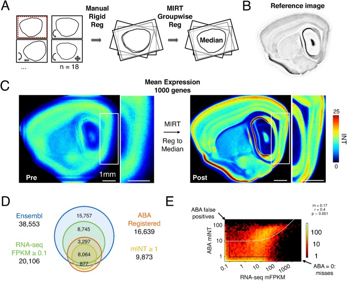
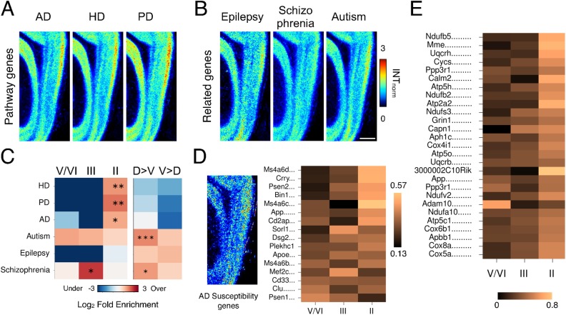
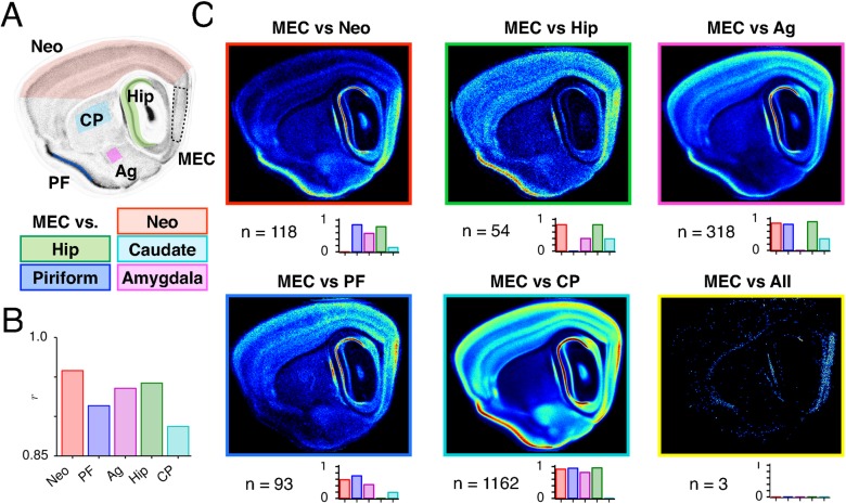
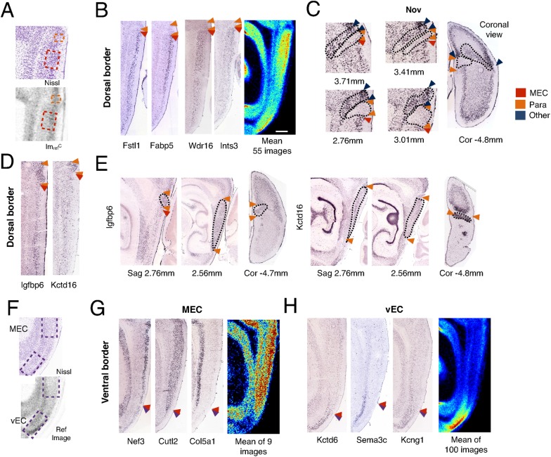
Neural circuits in the medial entorhinal cortex (MEC) encode an animal's position and orientation in space. Within the MEC spatial representations, including grid and directional firing fields, have a laminar and dorsoventral organization that corresponds to a similar topography of neuronal connectivity and cellular properties. Yet, in part due to the challenges of integrating anatomical data at the resolution of cortical layers and borders, we know little about the molecular components underlying this organization. To address this we develop a new computational pipeline for high-throughput analysis and comparison of in situ hybridization (ISH) images at laminar resolution. We apply this pipeline to ISH data for over 16,000 genes in the Allen Brain Atlas and validate our analysis with RNA sequencing of MEC tissue from adult mice. We find that differential gene expression delineates the borders of the MEC with neighboring brain structures and reveals its laminar and dorsoventral organization. We propose a new molecular basis for distinguishing the deep layers of the MEC and show that their similarity to corresponding layers of neocortex is greater than that of superficial layers. Our analysis identifies ion channel-, cell adhesion- and synapse-related genes as candidates for functional differentiation of MEC layers and for encoding of spatial information at different scales along the dorsoventral axis of the MEC. We also reveal laminar organization of genes related to disease pathology and suggest that a high metabolic demand predisposes layer II to neurodegenerative pathology. In principle, our computational pipeline can be applied to high-throughput analysis of many forms of neuroanatomical data. Our results support the hypothesis that differences in gene expression contribute to functional specialization of superficial layers of the MEC and dorsoventral organization of the scale of spatial representations.
内侧内嗅皮层(MEC)中的神经回路编码动物在空间中的位置和方向。在MEC的空间表征中,包括网格和方向发放场,具有层状和背腹组织,这与神经元连接和细胞特性的类似拓扑结构相对应。然而,部分由于在皮层层和边界分辨率下整合解剖数据的挑战,我们对这种组织的分子成分了解甚少。为了解决这个问题,我们开发了一种新的计算流程,用于在层状分辨率下对原位杂交(ISH)图像进行高通量分析和比较。我们将此流程应用于艾伦脑图谱中超过16000个基因的ISH数据,并用成年小鼠MEC组织的RNA测序验证了我们的分析。我们发现差异基因表达描绘了MEC与相邻脑结构的边界,并揭示了其层状和背腹组织。我们提出了一种区分MEC深层的新分子基础,并表明它们与新皮层相应层的相似性大于浅层。我们的分析确定了离子通道、细胞粘附和突触相关基因是MEC层功能分化以及沿MEC背腹轴在不同尺度上编码空间信息的候选基因。我们还揭示了与疾病病理学相关基因的层状组织,并表明高代谢需求使II层易患神经退行性病理学。原则上,我们的计算流程可应用于多种形式神经解剖学数据的高通量分析。我们的结果支持这样的假设,即基因表达差异有助于MEC浅层的功能特化以及空间表征尺度的背腹组织。