• 文献检索
  • 文档翻译
  • 深度研究
  • 学术资讯
  • Suppr Zotero 插件Zotero 插件
  • 邀请有礼
  • 套餐&价格
  • 历史记录
应用&插件
Suppr Zotero 插件Zotero 插件浏览器插件Mac 客户端Windows 客户端微信小程序
定价
高级版会员购买积分包购买API积分包
服务
文献检索文档翻译深度研究API 文档MCP 服务
关于我们
关于 Suppr公司介绍联系我们用户协议隐私条款
关注我们

Suppr 超能文献

核心技术专利:CN118964589B侵权必究
粤ICP备2023148730 号-1Suppr @ 2026

文献检索

告别复杂PubMed语法,用中文像聊天一样搜索,搜遍4000万医学文献。AI智能推荐,让科研检索更轻松。

立即免费搜索

文件翻译

保留排版,准确专业,支持PDF/Word/PPT等文件格式,支持 12+语言互译。

免费翻译文档

深度研究

AI帮你快速写综述,25分钟生成高质量综述,智能提取关键信息,辅助科研写作。

立即免费体验

临床照片中光化性角化病的自动检测。

Automated detection of actinic keratoses in clinical photographs.

作者信息

Hames Samuel C, Sinnya Sudipta, Tan Jean-Marie, Morze Conrad, Sahebian Azadeh, Soyer H Peter, Prow Tarl W

机构信息

Dermatology Research Center, School of Medicine, University of Queensland, Translational Research Institute, Princess Alexandra Hospital, Brisbane, Australia.

出版信息

PLoS One. 2015 Jan 23;10(1):e0112447. doi: 10.1371/journal.pone.0112447. eCollection 2015.

DOI:10.1371/journal.pone.0112447
PMID:25615930
原文链接:https://pmc.ncbi.nlm.nih.gov/articles/PMC4304708/
Abstract

BACKGROUND

Clinical diagnosis of actinic keratosis is known to have intra- and inter-observer variability, and there is currently no non-invasive and objective measure to diagnose these lesions.

OBJECTIVE

The aim of this pilot study was to determine if automatically detecting and circumscribing actinic keratoses in clinical photographs is feasible.

METHODS

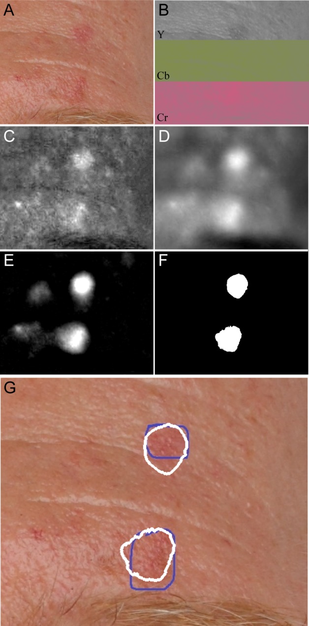
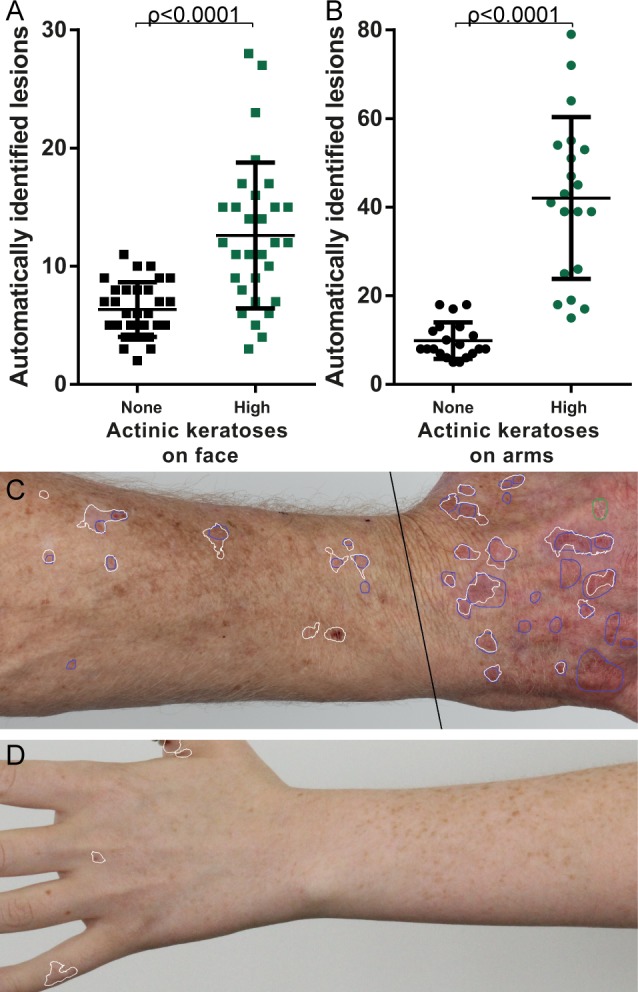
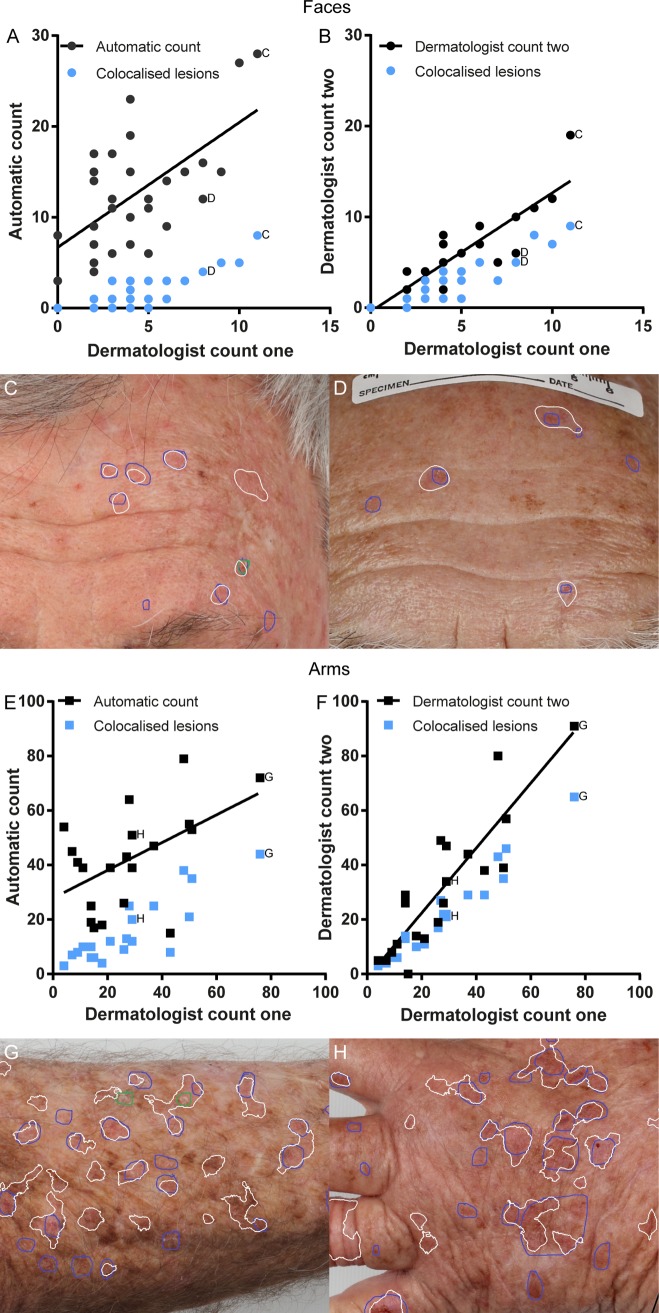
Photographs of the face and dorsal forearms were acquired in 20 volunteers from two groups: the first with at least on actinic keratosis present on the face and each arm, the second with no actinic keratoses. The photographs were automatically analysed using colour space transforms and morphological features to detect erythema. The automated output was compared with a senior consultant dermatologist's assessment of the photographs, including the intra-observer variability. Performance was assessed by the correlation between total lesions detected by automated method and dermatologist, and whether the individual lesions detected were in the same location as the dermatologist identified lesions. Additionally, the ability to limit false positives was assessed by automatic assessment of the photographs from the no actinic keratosis group in comparison to the high actinic keratosis group.

RESULTS

The correlation between the automatic and dermatologist counts was 0.62 on the face and 0.51 on the arms, compared to the dermatologist's intra-observer variation of 0.83 and 0.93 for the same. Sensitivity of automatic detection was 39.5% on the face, 53.1% on the arms. Positive predictive values were 13.9% on the face and 39.8% on the arms. Significantly more lesions (p<0.0001) were detected in the high actinic keratosis group compared to the no actinic keratosis group.

CONCLUSIONS

The proposed method was inferior to assessment by the dermatologist in terms of sensitivity and positive predictive value. However, this pilot study used only a single simple feature and was still able to achieve sensitivity of detection of 53.1% on the arms.This suggests that image analysis is a feasible avenue of investigation for overcoming variability in clinical assessment. Future studies should focus on more sophisticated features to improve sensitivity for actinic keratoses without erythema and limit false positives associated with the anatomical structures on the face.

摘要

背景

光化性角化病的临床诊断存在观察者内和观察者间的差异,目前尚无用于诊断这些病变的非侵入性客观方法。

目的

本初步研究的目的是确定在临床照片中自动检测和圈定光化性角化病是否可行。

方法

从两组20名志愿者处获取面部和前臂背部的照片:第一组面部和每只手臂上至少有一处光化性角化病,第二组没有光化性角化病。使用颜色空间变换和形态学特征对照片进行自动分析以检测红斑。将自动输出结果与皮肤科高级顾问对照片的评估进行比较,包括观察者内差异。通过自动方法检测到的总病变与皮肤科医生检测到的总病变之间的相关性以及检测到的单个病变是否与皮肤科医生确定的病变位于同一位置来评估性能。此外,通过与高光化性角化病组相比自动评估无光化性角化病组的照片来评估限制假阳性的能力。

结果

面部自动计数与皮肤科医生计数之间的相关性为0.62,手臂为0.51,而皮肤科医生的观察者内差异分别为0.83和0.93。面部自动检测的敏感性为39.5%,手臂为53.1%。面部的阳性预测值为13.9%,手臂为39.8%。高光化性角化病组检测到的病变明显多于无光化性角化病组(p<0.0001)。

结论

就敏感性和阳性预测值而言,所提出的方法不如皮肤科医生的评估。然而,这项初步研究仅使用了一个简单特征,仍能在手臂上实现53.1%的检测敏感性。这表明图像分析是克服临床评估差异的可行研究途径。未来的研究应侧重于更复杂的特征,以提高对无红斑光化性角化病的敏感性,并限制与面部解剖结构相关的假阳性。

https://cdn.ncbi.nlm.nih.gov/pmc/blobs/f5bb/4304708/6eb256cad4d6/pone.0112447.g004.jpg
https://cdn.ncbi.nlm.nih.gov/pmc/blobs/f5bb/4304708/d466988b280c/pone.0112447.g001.jpg
https://cdn.ncbi.nlm.nih.gov/pmc/blobs/f5bb/4304708/c34866a180ef/pone.0112447.g002.jpg
https://cdn.ncbi.nlm.nih.gov/pmc/blobs/f5bb/4304708/24c3f503c277/pone.0112447.g003.jpg
https://cdn.ncbi.nlm.nih.gov/pmc/blobs/f5bb/4304708/6eb256cad4d6/pone.0112447.g004.jpg

相似文献

1
Automated detection of actinic keratoses in clinical photographs.临床照片中光化性角化病的自动检测。
PLoS One. 2015 Jan 23;10(1):e0112447. doi: 10.1371/journal.pone.0112447. eCollection 2015.
2
The reliability of counting actinic keratosis.计数光化性角化病的可靠性。
Arch Dermatol Res. 2013 Nov;305(9):841-4. doi: 10.1007/s00403-013-1413-y. Epub 2013 Sep 18.
3
Imaging actinic keratosis by high-definition optical coherence tomography. Histomorphologic correlation: a pilot study.利用高清光学相干断层成像术对光化性角化病进行成像。组织形态学相关性:一项初步研究。
Exp Dermatol. 2013 Feb;22(2):93-7. doi: 10.1111/exd.12074. Epub 2013 Jan 10.
4
In vivo Confocal Laser Microscopy for monitoring of actinic keratosis treatment: a comparison with histopathologic assessment after treatment with topical 5% 5-fluorouracil.体内共聚焦激光显微镜在光化性角化病治疗监测中的应用:与外用 5% 5-氟尿嘧啶治疗后组织病理学评估的比较。
J Eur Acad Dermatol Venereol. 2018 Jul;32(7):1155-1163. doi: 10.1111/jdv.14716. Epub 2018 Jan 23.
5
Assessing the Concordance of Actinic Keratosis Counts on Digital Photographs with Clinical Examination in Organ Transplant Recipients.评估器官移植受者数字照片中光化性角化病计数与临床检查结果的一致性。
Acta Derm Venereol. 2017 Mar 10;97(3):351-353. doi: 10.2340/00015555-2539.
6
Primary care clinicians' performance for detecting actinic keratoses and skin cancer.基层医疗临床医生在检测光化性角化病和皮肤癌方面的表现。
Arch Intern Med. 1997 May 12;157(9):985-90.
7
Imiquimod 3.75% for field-directed therapy of actinic keratosis: results of a prospective case-series study in Greece.咪喹莫特 3.75% 用于光化性角化病的定向治疗:希腊前瞻性病例系列研究结果。
Int J Dermatol. 2019 Sep;58(9):1040-1044. doi: 10.1111/ijd.14397. Epub 2019 Feb 19.
8
Role of In Vivo Reflectance Confocal Microscopy in the Analysis of Melanocytic Lesions.体内反射共聚焦显微镜在黑素细胞性病变分析中的作用
Acta Dermatovenerol Croat. 2018 Apr;26(1):64-67.
9
Inner gray halo, a novel dermoscopic feature for the diagnosis of pigmented actinic keratosis: clues for the differential diagnosis with lentigo maligna.内灰色晕环,一种用于诊断色素性光化性角化病的新型皮肤镜特征:与恶性雀斑样痣鉴别诊断的线索。
J Am Acad Dermatol. 2014 Oct;71(4):708-15. doi: 10.1016/j.jaad.2014.05.025. Epub 2014 Jun 17.
10
Early clinical response to 5-fluorouracil 0.5% and salicylic acid 10% topical solution in the treatment of actinic keratoses of the head: an observational study.5-氟尿嘧啶0.5%与水杨酸10%外用溶液治疗头部光化性角化病的早期临床反应:一项观察性研究。
J Dermatolog Treat. 2022 Aug;33(5):2664-2669. doi: 10.1080/09546634.2022.2067817. Epub 2022 May 26.

引用本文的文献

1
AΚtransU-Net: Transformer-Equipped U-Net Model for Improved Actinic Keratosis Detection in Clinical Photography.AΚtransU-Net:配备Transformer的U-Net模型用于改善临床摄影中光化性角化病的检测
Diagnostics (Basel). 2025 Jul 10;15(14):1752. doi: 10.3390/diagnostics15141752.
2
Insights, Advantages, and Barriers of Teledermatology vs. Face-to-Face Dermatology for the Diagnosis and Follow-Up of Non-Melanoma Skin Cancer: A Systematic Review.远程皮肤病学与面对面皮肤病学在非黑色素瘤皮肤癌诊断和随访中的见解、优势及障碍:一项系统综述
Cancers (Basel). 2024 Jan 30;16(3):578. doi: 10.3390/cancers16030578.
3
Where Artificial Intelligence Can Take Us in the Management and Understanding of Cancerization Fields.

本文引用的文献

1
Improvement in precision of counting actinic keratoses.光化性角化病计数精度的提高。
Br J Dermatol. 2014 Jan;170(1):188-91. doi: 10.1111/bjd.12629.
2
The reliability of counting actinic keratosis.计数光化性角化病的可靠性。
Arch Dermatol Res. 2013 Nov;305(9):841-4. doi: 10.1007/s00403-013-1413-y. Epub 2013 Sep 18.
3
Reliability of quantification measures of actinic keratosis.光化性角化病定量测量指标的可靠性。
人工智能在癌症化领域的管理与理解方面能引领我们走向何方。
Cancers (Basel). 2023 Nov 2;15(21):5264. doi: 10.3390/cancers15215264.
4
The Promise of Semantic Segmentation in Detecting Actinic Keratosis Using Clinical Photography in the Wild.在自然环境下使用临床摄影检测光化性角化病中语义分割的前景。
Cancers (Basel). 2023 Oct 5;15(19):4861. doi: 10.3390/cancers15194861.
5
Inter- and Intra-physician variation in quantifying actinic keratosis skin photodamage.医生之间和医生内部在量化光化性角化病皮肤光损伤方面的差异。
J Clin Investig Dermatol. 2020 Sep;8(2). Epub 2020 Sep 6.
6
Vascular patterns in basal cell carcinoma: Dermoscopic, confocal and histopathological perspectives.基底细胞癌的血管形态:皮肤镜、共聚焦显微镜及组织病理学视角
Oncol Lett. 2019 May;17(5):4112-4125. doi: 10.3892/ol.2019.10070. Epub 2019 Feb 25.
7
A Computer-Aided Decision Support System for Detection and Localization of Cutaneous Vasculature in Dermoscopy Images Via Deep Feature Learning.基于深度特征学习的皮肤镜图像皮肤血管检测与定位的计算机辅助决策支持系统。
J Med Syst. 2018 Jan 9;42(2):33. doi: 10.1007/s10916-017-0885-2.
Br J Dermatol. 2013 Dec;169(6):1219-22. doi: 10.1111/bjd.12591.
4
Guided image filtering.引导图像滤波。
IEEE Trans Pattern Anal Mach Intell. 2013 Jun;35(6):1397-409. doi: 10.1109/TPAMI.2012.213.
5
Therapeutic strategies for actinic keratoses--a systematic review.光化性角化病的治疗策略——系统评价。
Eur J Dermatol. 2013 Jan-Feb;23(1):14-32. doi: 10.1684/ejd.2013.1923.
6
A system for the detection of pigment network in dermoscopy images using directional filters.基于方向滤波器的皮肤镜图像色素网络检测系统。
IEEE Trans Biomed Eng. 2012 Oct;59(10):2744-54. doi: 10.1109/TBME.2012.2209423. Epub 2012 Jul 18.
7
Dermoscopic diagnosis of melanoma in a 4D space constructed by active contour extracted features.基于主动轮廓提取特征的 4D 空间中黑色素瘤的皮肤镜诊断。
Comput Med Imaging Graph. 2012 Oct;36(7):572-9. doi: 10.1016/j.compmedimag.2012.06.002. Epub 2012 Jul 18.
8
Lesion border detection in dermoscopy images using ensembles of thresholding methods.利用阈值方法集进行皮肤镜图像的病灶边界检测。
Skin Res Technol. 2013 Feb;19(1):e252-8. doi: 10.1111/j.1600-0846.2012.00636.x. Epub 2012 Jun 7.
9
Automatic telangiectasia analysis in dermoscopy images using adaptive critic design.利用自适应评价设计进行皮肤镜图像中的自动毛细血管扩张分析。
Skin Res Technol. 2012 Nov;18(4):389-96. doi: 10.1111/j.1600-0846.2011.00584.x. Epub 2011 Dec 5.
10
Three-phase general border detection method for dermoscopy images using non-uniform illumination correction.基于非均匀光照校正的皮肤镜图像三相广义边界检测方法
Skin Res Technol. 2012 Aug;18(3):290-300. doi: 10.1111/j.1600-0846.2011.00569.x. Epub 2011 Sep 6.