Wang Yong, Zhang Hui, Wang Zhe, Geng Zuojun, Liu Huaijun, Yang Haiqing, Song Peng, Liu Qing
Department of Radiology, the Second Hospital of Hebei Medical University, Shijiazhuang 050000, Hebei Province, China.
Department of Radiology, Hebei General Hospital, Shijiazhuang 050051, Hebei Province, China.
Neural Regen Res. 2012 Aug 25;7(24):1873-80. doi: 10.3969/j.issn.1673-5374.2012.24.005.
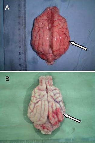
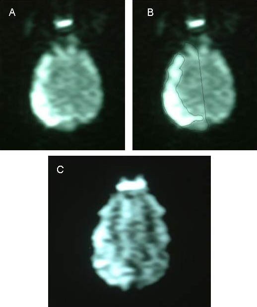
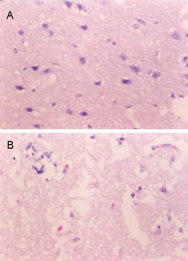
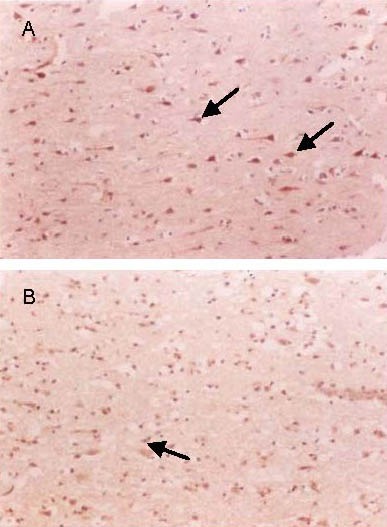
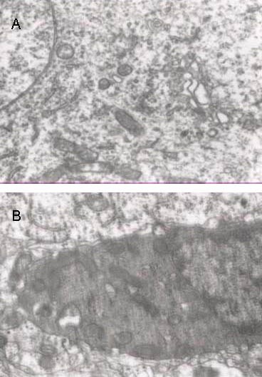
A model of focal cerebral ischemic infarction was established in dogs through middle cerebral artery occlusion of the right side. Thirty minutes after occlusion, models were injected with nerve growth factor adjacent to the infarct locus. The therapeutic effect of nerve growth factor against cerebral infarction was assessed using the hemisphere anomalous volume ratio, a quantitative index of diffusion-weighted MRI. At 6 hours, 24 hours, 7 days and 3 months after modeling, the hemisphere anomalous volume ratio was significantly reduced after treatment with nerve growth factor. Hematoxylin-eosin staining, immunohistochemistry, electron microscopy and neurological function scores showed that infarct defects were slightly reduced and neurological function significantly improved after nerve growth factor treatment. This result was consistent with diffusion-weighted MRI measurements. Experimental findings indicate that nerve growth factor can protect against cerebral infarction, and that the hemisphere anomalous volume ratio of diffusion-weighted MRI can be used to evaluate the therapeutic effect.
通过右侧大脑中动脉闭塞在犬类中建立局灶性脑缺血梗死模型。闭塞30分钟后,在梗死灶附近注射神经生长因子。使用扩散加权磁共振成像的定量指标——半球异常体积比来评估神经生长因子对脑梗死的治疗效果。建模后6小时、24小时、7天和3个月时,神经生长因子治疗后半球异常体积比显著降低。苏木精-伊红染色、免疫组织化学、电子显微镜检查和神经功能评分显示,神经生长因子治疗后梗死缺损略有减少,神经功能显著改善。这一结果与扩散加权磁共振成像测量结果一致。实验结果表明,神经生长因子可预防脑梗死,且扩散加权磁共振成像的半球异常体积比可用于评估治疗效果。