Sawant Anuradha, House Andrew A, Chesworth Bert M, Connelly Denise M, Lindsay Robert, Gati Joe, Bartha Robert, Overend Tom J
Western University, London, Ontario, Canada London Health Sciences Center, University Hospital Campus, London, Ontario, Canada.
Division of Nephrology, London Health Sciences Centre, Western University, London, Ontario, Canada.
Physiol Rep. 2015 Jan 27;3(1). doi: 10.14814/phy2.12219. Print 2015 Jan 1.
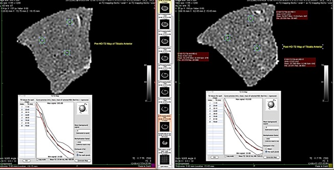
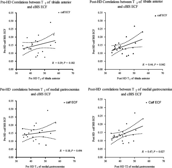
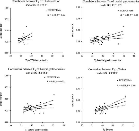
Establishing the effect of fluctuating extracellular fluid (ECF) volume on muscle strength in people with end-stage renal disease (ESRD) on hemodialysis (HD) is essential, as inadequate hydration of the skeletal muscles impacts its strength and endurance. Bioelectrical impedance spectroscopy (BIS) has been a widely used method for estimating ECF volume of a limb or calf segment. Magnetic resonance imaging (MRI)-acquired transverse relaxation times (T2) has also been used for estimating ECF volumes of individual skeletal muscles. The purpose of this study was to determine the association between T2 (gold standard) of tibialis anterior (TA), medial (MG), and lateral gastrocnemius (LG), and soleus muscles and calf BIS ECF, in healthy and in people with ESRD/HD. Calf BIS and MRI measures were collected on two occasions before and after HD session in people with ESRD/HD and on a single occasion for the healthy participants. Linear regression analysis was used to establish the association between these measures. Thirty-two healthy and 22 participants on HD were recruited. The association between T2 of TA, LG, MG, and soleus muscles and ratio of calf BIS-acquired ECF and intracellular fluids (ICF) were: TA: β = 0.30, P > 0.05; LG: β = 0.37, P = 0.035; MG: β = 0.43, P = 0.014; soleus: β = 0.60, P < 0.001. For the HD group, calf ECF was significantly associated with T2 of TA (β = 0.44, P = 0.042), and medial gastrocnemius (β = 0.47, P = 0.027) following HD only. Hence BIS-acquired measures cannot be used to measure ECF volumes of a single muscle in the ESRD/HD population; however, BIS could be utilized to estimate ratio of ECF: ICF in healthy population for the LG, MG, and soleus muscles.
确定终末期肾病(ESRD)患者接受血液透析(HD)时,细胞外液(ECF)容量波动对肌肉力量的影响至关重要,因为骨骼肌水合不足会影响其力量和耐力。生物电阻抗光谱法(BIS)一直是用于估计肢体或小腿段ECF容量的广泛使用的方法。磁共振成像(MRI)获取的横向弛豫时间(T2)也已用于估计单个骨骼肌的ECF容量。本研究的目的是确定健康人群以及ESRD/HD患者中,胫骨前肌(TA)、内侧腓肠肌(MG)、外侧腓肠肌(LG)和比目鱼肌的T2(金标准)与小腿BIS ECF之间的关联。在ESRD/HD患者的HD治疗前后两次收集小腿BIS和MRI测量值,而健康参与者仅收集一次。采用线性回归分析来确定这些测量值之间的关联。招募了32名健康参与者和22名接受HD治疗的参与者。TA、LG、MG和比目鱼肌的T2与小腿BIS获取的ECF和细胞内液(ICF)的比率之间的关联为:TA:β = 0.30,P > 0.05;LG:β = 0.37,P = 0.035;MG:β = 0.43,P = 0.014;比目鱼肌:β = 0.60,P < 0.001。对于HD组,仅在HD治疗后,小腿ECF与TA的T2(β = 0.44,P = 0.042)和内侧腓肠肌的T2(β = 0.47,P = 0.027)显著相关。因此,在ESRD/HD人群中,BIS获取的测量值不能用于测量单个肌肉的ECF容量;然而,BIS可用于估计健康人群中LG、MG和比目鱼肌的ECF:ICF比率。