• 文献检索
  • 文档翻译
  • 深度研究
  • 学术资讯
  • Suppr Zotero 插件Zotero 插件
  • 邀请有礼
  • 套餐&价格
  • 历史记录
应用&插件
Suppr Zotero 插件Zotero 插件浏览器插件Mac 客户端Windows 客户端微信小程序
定价
高级版会员购买积分包购买API积分包
服务
文献检索文档翻译深度研究API 文档MCP 服务
关于我们
关于 Suppr公司介绍联系我们用户协议隐私条款
关注我们

Suppr 超能文献

核心技术专利:CN118964589B侵权必究
粤ICP备2023148730 号-1Suppr @ 2026

文献检索

告别复杂PubMed语法,用中文像聊天一样搜索,搜遍4000万医学文献。AI智能推荐,让科研检索更轻松。

立即免费搜索

文件翻译

保留排版,准确专业,支持PDF/Word/PPT等文件格式,支持 12+语言互译。

免费翻译文档

深度研究

AI帮你快速写综述,25分钟生成高质量综述,智能提取关键信息,辅助科研写作。

立即免费体验

相似文献

1
Automated analysis of cell-matrix adhesions in 2D and 3D environments.二维和三维环境中细胞-基质黏附的自动化分析。
Sci Rep. 2015 Jan 29;5:8124. doi: 10.1038/srep08124.
2
Taking cell-matrix adhesions to the third dimension.将细胞-基质黏附拓展到三维空间。
Science. 2001 Nov 23;294(5547):1708-12. doi: 10.1126/science.1064829.
3
Cell type-specific adaptation of cellular and nuclear volume in micro-engineered 3D environments.微工程 3D 环境中细胞和核体积的细胞类型特异性适应性。
Biomaterials. 2015 Nov;69:121-32. doi: 10.1016/j.biomaterials.2015.08.016. Epub 2015 Aug 8.
4
Do cancer cells have distinct adhesions in 3D collagen matrices and in vivo?癌细胞在 3D 胶原基质中和体内是否具有独特的黏附性?
Eur J Cell Biol. 2012 Nov-Dec;91(11-12):930-7. doi: 10.1016/j.ejcb.2012.07.005. Epub 2012 Aug 29.
5
Automated tracking of unmarked cells migrating in three-dimensional matrices applied to anti-cancer drug screening.自动化追踪三维基质中无标记细胞的迁移,应用于抗癌药物筛选。
Exp Cell Res. 2010 Jan 15;316(2):181-93. doi: 10.1016/j.yexcr.2009.10.004. Epub 2009 Oct 14.
6
Imaging cells within 3D cell-derived matrix.对三维细胞衍生基质中的细胞进行成像。
Methods Mol Biol. 2011;769:53-64. doi: 10.1007/978-1-61779-207-6_5.
7
Disentangling Membrane Dynamics and Cell Migration; Differential Influences of F-actin and Cell-Matrix Adhesions.解析膜动力学与细胞迁移;F-肌动蛋白和细胞-基质黏附的不同影响
PLoS One. 2015 Aug 6;10(8):e0135204. doi: 10.1371/journal.pone.0135204. eCollection 2015.
8
Cell-matrix adhesions in 3D.三维细胞-基质黏附。
Matrix Biol. 2011 Sep;30(7-8):363-8. doi: 10.1016/j.matbio.2011.06.001. Epub 2011 Jun 23.
9
Quantification of collagen contraction in three-dimensional cell culture.三维细胞培养中胶原蛋白收缩的定量分析。
Methods Cell Biol. 2015;125:353-72. doi: 10.1016/bs.mcb.2014.10.017. Epub 2015 Jan 8.
10
Podosome-type adhesions and focal adhesions, so alike yet so different.足体样黏附与黏着斑,如此相似却又如此不同。
Eur J Cell Biol. 2008 Sep;87(8-9):491-506. doi: 10.1016/j.ejcb.2008.02.012. Epub 2008 Apr 15.

引用本文的文献

1
Enhanced statistical sampling reveals microscopic complexity in the talin mechanosensor folding energy landscape.增强的统计抽样揭示了踝蛋白机械传感器折叠能量景观中的微观复杂性。
Nat Phys. 2023 Jan;19(1):52-60. doi: 10.1038/s41567-022-01808-4. Epub 2022 Nov 7.
2
Meaningful connections: Interrogating the role of physical fibroblast cell-cell communication in cancer.有意义的连接:探究物理成纤维细胞细胞间通讯在癌症中的作用。
Adv Cancer Res. 2022;154:141-168. doi: 10.1016/bs.acr.2022.01.004. Epub 2022 Feb 24.
3
A Focal Adhesion Filament Cross-correlation Kit for fast, automated segmentation and correlation of focal adhesions and actin stress fibers in cells.用于快速、自动化分割和关联细胞中的焦点黏附及其肌动蛋白应力纤维的焦点黏附丝共相关试剂盒。
PLoS One. 2021 Sep 10;16(9):e0250749. doi: 10.1371/journal.pone.0250749. eCollection 2021.
4
Wavelet Imaging on Multiple Scales (WIMS) reveals focal adhesion distributions, dynamics and coupling between actomyosin bundle stability.多尺度小波成像(WIMS)揭示了粘着斑分布、动力学以及肌动球蛋白束稳定性之间的耦合关系。
PLoS One. 2017 Oct 19;12(10):e0186058. doi: 10.1371/journal.pone.0186058. eCollection 2017.
5
Mechanosensing via cell-matrix adhesions in 3D microenvironments.三维微环境中通过细胞-基质黏附进行的机械传感
Exp Cell Res. 2016 Apr 10;343(1):60-66. doi: 10.1016/j.yexcr.2015.10.033. Epub 2015 Nov 6.

本文引用的文献

1
Activation of Rac by Asef2 promotes myosin II-dependent contractility to inhibit cell migration on type I collagen.Asef2介导的Rac激活增强肌球蛋白II依赖性收缩能力,从而抑制细胞在I型胶原上的迁移。
J Cell Sci. 2013 Dec 15;126(Pt 24):5585-97. doi: 10.1242/jcs.131060. Epub 2013 Oct 21.
2
Matrix microarchitecture and myosin II determine adhesion in 3D matrices.基质微架构和肌球蛋白 II 决定 3D 基质中的黏附。
Curr Biol. 2013 Sep 9;23(17):1607-19. doi: 10.1016/j.cub.2013.06.053. Epub 2013 Aug 8.
3
Dynamic regulation of the structure and functions of integrin adhesions.整合素黏附结构与功能的动态调控。
Dev Cell. 2013 Mar 11;24(5):447-58. doi: 10.1016/j.devcel.2013.02.012.
4
The origins of the molecular era of adhesion research.分子时代黏附研究的起源。
Nat Rev Mol Cell Biol. 2012 Dec;13(12):805-11. doi: 10.1038/nrm3473. Epub 2012 Nov 15.
5
Do cancer cells have distinct adhesions in 3D collagen matrices and in vivo?癌细胞在 3D 胶原基质中和体内是否具有独特的黏附性?
Eur J Cell Biol. 2012 Nov-Dec;91(11-12):930-7. doi: 10.1016/j.ejcb.2012.07.005. Epub 2012 Aug 29.
6
Assembly and disassembly of cell matrix adhesions.细胞基质黏附的组装与拆卸。
Curr Opin Cell Biol. 2012 Oct;24(5):569-81. doi: 10.1016/j.ceb.2012.06.010. Epub 2012 Jul 19.
7
The endosomal adaptor protein APPL1 impairs the turnover of leading edge adhesions to regulate cell migration.内体衔接蛋白 APPL1 损害前沿黏附的周转率,从而调节细胞迁移。
Mol Biol Cell. 2012 Apr;23(8):1486-99. doi: 10.1091/mbc.E11-02-0124. Epub 2012 Feb 29.
8
Micro-environmental control of cell migration--myosin IIA is required for efficient migration in fibrillar environments through control of cell adhesion dynamics.细胞迁移的微环境控制——肌球蛋白 IIA 通过控制细胞黏附动力学,对于在纤维环境中的有效迁移是必需的。
J Cell Sci. 2012 May 1;125(Pt 9):2244-56. doi: 10.1242/jcs.098806. Epub 2012 Feb 10.
9
The structure of cell-matrix adhesions: the new frontier.细胞基质黏附的结构:新前沿。
Curr Opin Cell Biol. 2012 Feb;24(1):134-40. doi: 10.1016/j.ceb.2011.12.001. Epub 2011 Dec 22.
10
Adhesion dynamics at a glance.粘连动力学一览。
J Cell Sci. 2011 Dec 1;124(Pt 23):3923-7. doi: 10.1242/jcs.095653.

二维和三维环境中细胞-基质黏附的自动化分析。

Automated analysis of cell-matrix adhesions in 2D and 3D environments.

作者信息

Broussard Joshua A, Diggins Nicole L, Hummel Stephen, Georgescu Walter, Quaranta Vito, Webb Donna J

机构信息

Department of Biological Sciences and Vanderbilt Kennedy Center for Research on Human Development, Vanderbilt University, Nashville, Tennessee 37235.

Center for Cancer Systems Biology at Vanderbilt, Vanderbilt University, Nashville, Tennessee 37235.

出版信息

Sci Rep. 2015 Jan 29;5:8124. doi: 10.1038/srep08124.

DOI:10.1038/srep08124
PMID:25630460
原文链接:https://pmc.ncbi.nlm.nih.gov/articles/PMC4309964/
Abstract

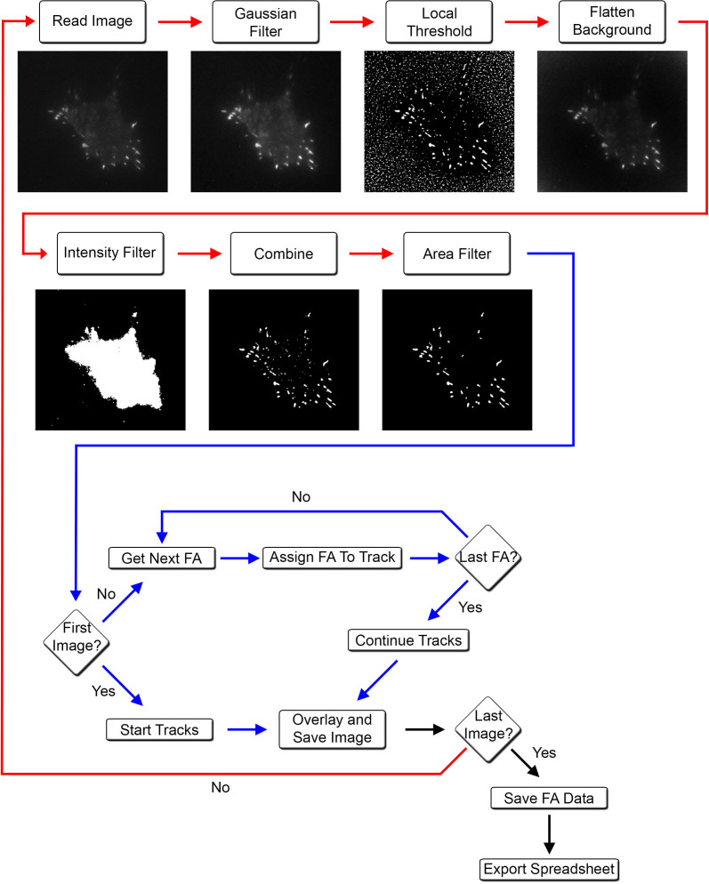
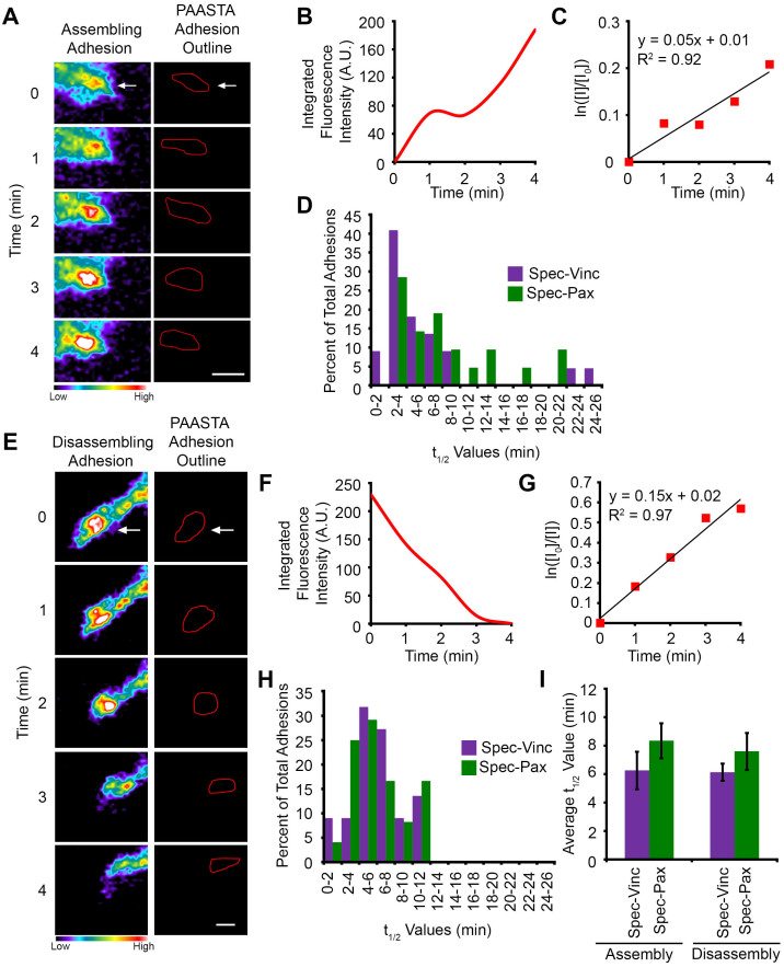
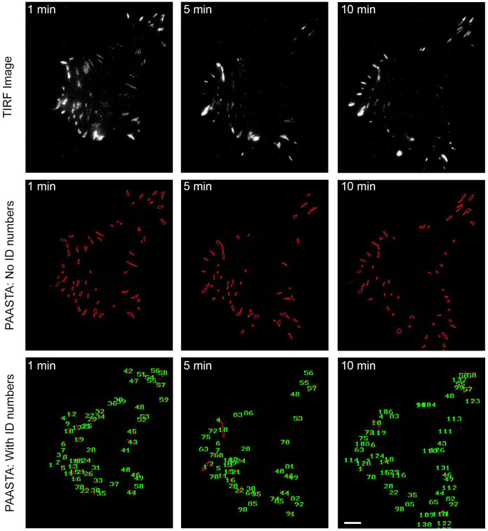
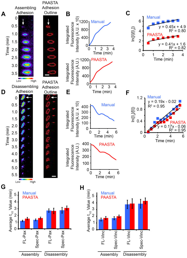
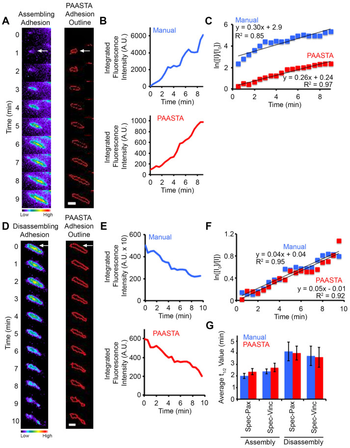
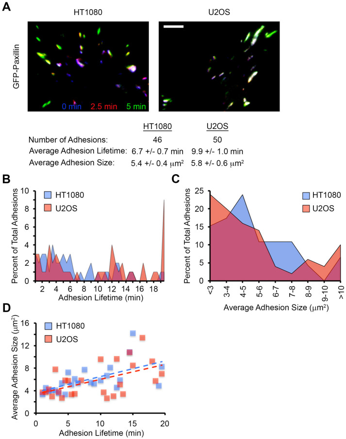
Cell-matrix adhesions are of great interest because of their contribution to numerous biological processes, including cell migration, differentiation, proliferation, survival, tissue morphogenesis, wound healing, and tumorigenesis. Adhesions are dynamic structures that are classically defined on two-dimensional (2D) substrates, though the need to analyze adhesions in more physiologic three-dimensional (3D) environments is being increasingly recognized. However, progress has been greatly hampered by the lack of available tools to analyze adhesions in 3D environments. To address this need, we have developed a platform for the automated analysis, segmentation, and tracking of adhesions (PAASTA) based on an open source MATLAB framework, CellAnimation. PAASTA enables the rapid analysis of adhesion dynamics and many other adhesion characteristics, such as lifetime, size, and location, in 3D environments and on traditional 2D substrates. We manually validate PAASTA and utilize it to quantify rate constants for adhesion assembly and disassembly as well as adhesion lifetime and size in 3D matrices. PAASTA will be a valuable tool for characterizing adhesions and for deciphering the molecular mechanisms that regulate adhesion dynamics in 3D environments.

摘要

细胞与基质的黏附备受关注,因为它们对众多生物学过程有重要作用,包括细胞迁移、分化、增殖、存活、组织形态发生、伤口愈合和肿瘤发生。黏附是动态结构,传统上是在二维(2D)基质上定义的,不过人们越来越认识到需要在更接近生理状态的三维(3D)环境中分析黏附。然而,由于缺乏在3D环境中分析黏附的可用工具,进展受到了极大阻碍。为满足这一需求,我们基于开源MATLAB框架CellAnimation开发了一个用于黏附自动分析、分割和跟踪的平台(PAASTA)。PAASTA能够在3D环境和传统2D基质上快速分析黏附动力学以及许多其他黏附特性,如寿命、大小和位置。我们手动验证了PAASTA,并利用它来量化3D基质中黏附组装和解聚的速率常数以及黏附寿命和大小。PAASTA将成为表征黏附以及解读在3D环境中调节黏附动力学的分子机制的宝贵工具。

https://cdn.ncbi.nlm.nih.gov/pmc/blobs/6166/4309964/b2e3007bbe06/srep08124-f6.jpg
https://cdn.ncbi.nlm.nih.gov/pmc/blobs/6166/4309964/10d9b3e8ccfe/srep08124-f1.jpg
https://cdn.ncbi.nlm.nih.gov/pmc/blobs/6166/4309964/b942453a71ca/srep08124-f2.jpg
https://cdn.ncbi.nlm.nih.gov/pmc/blobs/6166/4309964/0a42727f142d/srep08124-f3.jpg
https://cdn.ncbi.nlm.nih.gov/pmc/blobs/6166/4309964/fe2eb2b811ac/srep08124-f4.jpg
https://cdn.ncbi.nlm.nih.gov/pmc/blobs/6166/4309964/4b8100f6404b/srep08124-f5.jpg
https://cdn.ncbi.nlm.nih.gov/pmc/blobs/6166/4309964/b2e3007bbe06/srep08124-f6.jpg