Yan Gen, Dai Zhuozhi, Xuan Yinghua, Wu Renhua
Department of Radiology, Affiliated Hospital, Jiangnan University, Wuxi, Jiangsu 214062, P.R. China.
Department of Radiology, The Second Affiliated Hospital, Shantou University Medical College, Shantou, Guangdong 515041, P.R. China.
Mol Med Rep. 2015 Jun;11(6):4109-14. doi: 10.3892/mmr.2015.3283. Epub 2015 Jan 30.
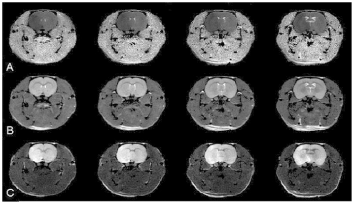
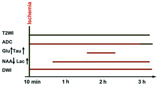
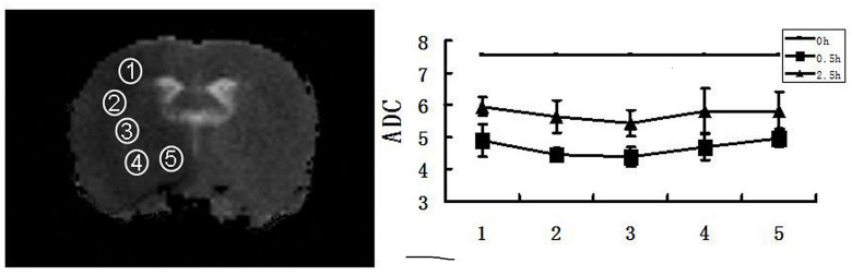
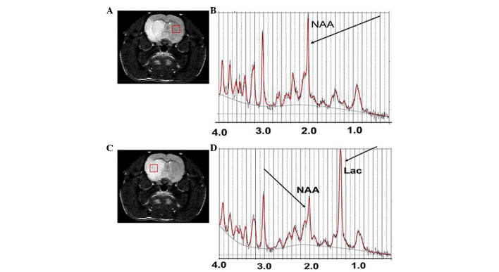
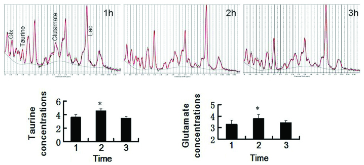
Despite improvements in imaging techniques, it remains challenging to quantitatively assess the time of ischemic onset of an acute ischemic stroke. It is crucial to evaluate the early signs of infarction, which are predictive of responses to recombinant tissue plasminogen activator within a treatment window of 4.5 h after stroke induction. The aim of the present study was to assess and quantify the onset time for hyperacute middle cerebral artery occlusion (MCAO) ischemic stroke by measuring the apparent diffusion coefficient (ADC) of diffusion‑weighted imaging (DWI) and 1H‑magnetic resonance spectroscopy (MRS) at 7.0 T. DWI, conventional T2‑weighted imaging (T2WI) and subsequent focal ADCs were employed to evaluate ischemic brain lesions in a rat model of MCAO (n=20) at different time‑points following a stroke. A quantitation of local changes in metabolite concentrations within the lesions was performed using MRS. Proton metabolites were quantified automatically using LCModel software. At 30 min after MCAO, intense signals were observed in the DWI spectra of all animals. No abnormal signal was observed within 3 h by T2WI. ADC images of the central area, peripheral striping and on the fringes of the infarction demonstrated a lower signal than that of the normal side. The ADC decreased significantly within 30 min after infarction, followed by a gradual elevation in volatility levels and then becoming relatively stable at a lower level 3 h later. MRS exhibited a consistent elevation of lactate and reduced N‑acetyl aspartic acid. Glutamate and taurine reached a maximum 2 h after MCAO and began to decrease 1 h later. In conclusion, the present study demonstrated that hyperacute ischemic stroke can be quantitatively detected with the application of ADC, DWI and MRS. These methods may also be used to quantitatively assess the ischemic onset time of a hyperacute stroke.
尽管成像技术有所改进,但定量评估急性缺血性中风的缺血发作时间仍然具有挑战性。评估梗死的早期迹象至关重要,这些迹象可预测在中风诱导后4.5小时的治疗窗口内对重组组织型纤溶酶原激活剂的反应。本研究的目的是通过在7.0T下测量扩散加权成像(DWI)的表观扩散系数(ADC)和1H磁共振波谱(MRS)来评估和量化超急性大脑中动脉闭塞(MCAO)缺血性中风的发作时间。在中风后的不同时间点,采用DWI、传统T2加权成像(T2WI)和随后的局灶性ADC来评估MCAO大鼠模型(n = 20)中的缺血性脑损伤。使用MRS对病变内代谢物浓度的局部变化进行定量。使用LCModel软件自动对质子代谢物进行定量。MCAO后30分钟,在所有动物的DWI光谱中观察到强烈信号。T2WI在3小时内未观察到异常信号。梗死中心区域、外周条纹和边缘的ADC图像显示信号低于正常侧。梗死30分钟内ADC显著下降,随后波动性水平逐渐升高,3小时后在较低水平相对稳定。MRS显示乳酸持续升高,N-乙酰天门冬氨酸减少。谷氨酸和牛磺酸在MCAO后2小时达到最大值,1小时后开始下降。总之,本研究表明,应用ADC、DWI和MRS可以定量检测超急性缺血性中风。这些方法也可用于定量评估超急性中风的缺血发作时间。