Jiménez-Xarrié Elena, Davila Myriam, Candiota Ana Paula, Delgado-Mederos Raquel, Ortega-Martorell Sandra, Julià-Sapé Margarida, Arús Carles, Martí-Fàbregas Joan
Stroke Unit, Department of Neurology, Hospital de la Santa Creu i Sant Pau, IIB-Sant Pau, Sant Antoni Maria Claret 167, 08025, Barcelona, Spain.
Departament de Bioquímica i Biologia Molecular, Unitat de Biociències, Edifici C, Universitat Autònoma de Barcelona, 08193, Cerdanyola del Vallès, Spain.
BMC Neurosci. 2017 Jan 13;18(1):13. doi: 10.1186/s12868-016-0328-x.
Magnetic resonance spectroscopy (MRS) provides non-invasive information about the metabolic pattern of the brain parenchyma in vivo. The SpectraClassifier software performs MRS pattern-recognition by determining the spectral features (metabolites) which can be used objectively to classify spectra. Our aim was to develop an Infarct Evolution Classifier and a Brain Regions Classifier in a rat model of focal ischemic stroke using SpectraClassifier.
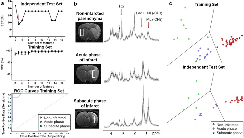
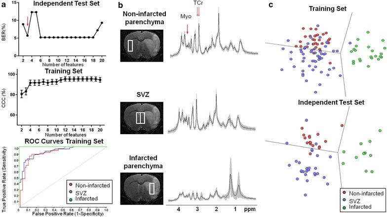
A total of 164 single-voxel proton spectra obtained with a 7 Tesla magnet at an echo time of 12 ms from non-infarcted parenchyma, subventricular zones and infarcted parenchyma were analyzed with SpectraClassifier ( http://gabrmn.uab.es/?q=sc ). The spectra corresponded to Sprague-Dawley rats (healthy rats, n = 7) and stroke rats at day 1 post-stroke (acute phase, n = 6 rats) and at days 7 ± 1 post-stroke (subacute phase, n = 14). In the Infarct Evolution Classifier, spectral features contributed by lactate + mobile lipids (1.33 ppm), total creatine (3.05 ppm) and mobile lipids (0.85 ppm) distinguished among non-infarcted parenchyma (100% sensitivity and 100% specificity), acute phase of infarct (100% sensitivity and 95% specificity) and subacute phase of infarct (78% sensitivity and 100% specificity). In the Brain Regions Classifier, spectral features contributed by myoinositol (3.62 ppm) and total creatine (3.04/3.05 ppm) distinguished among infarcted parenchyma (100% sensitivity and 98% specificity), non-infarcted parenchyma (84% sensitivity and 84% specificity) and subventricular zones (76% sensitivity and 93% specificity).
SpectraClassifier identified candidate biomarkers for infarct evolution (mobile lipids accumulation) and different brain regions (myoinositol content).
磁共振波谱(MRS)可在体内提供有关脑实质代谢模式的非侵入性信息。SpectraClassifier软件通过确定可用于客观分类波谱的光谱特征(代谢物)来执行MRS模式识别。我们的目的是使用SpectraClassifier在局灶性缺血性中风大鼠模型中开发梗死演变分类器和脑区分类器。
使用SpectraClassifier(http://gabrmn.uab.es/?q=sc)分析了在回波时间为12毫秒时用7特斯拉磁体从非梗死脑实质、脑室下区和梗死脑实质获得的总共164个单体素质子波谱。这些波谱对应于Sprague-Dawley大鼠(健康大鼠,n = 7)以及中风后第1天(急性期,n = 6只大鼠)和中风后7±1天(亚急性期,n = 14)的中风大鼠。在梗死演变分类器中,乳酸+流动脂质(1.33 ppm)、总肌酸(3.05 ppm)和流动脂质(0.85 ppm)贡献的光谱特征可区分非梗死脑实质(敏感性100%,特异性100%)、梗死急性期(敏感性100%,特异性95%)和梗死亚急性期(敏感性78%,特异性100%)。在脑区分类器中,肌醇(3.62 ppm)和总肌酸(3.04/3.05 ppm)贡献的光谱特征可区分梗死脑实质(敏感性100%,特异性98%)、非梗死脑实质(敏感性84%,特异性84%)和脑室下区(敏感性76%,特异性93%)。
SpectraClassifier识别出了梗死演变(流动脂质积累)和不同脑区(肌醇含量)的候选生物标志物。