Osborne Andrew, Aldarwesh Amal, Rhodes Jeremy D, Broadway David C, Everitt Claire, Sanderson Julie
School of Pharmacy, University of East Anglia, Norwich, United Kingdom.
School of Biological Sciences, University of East Anglia, Norwich, United Kingdom.
PLoS One. 2015 Jan 30;10(1):e0115591. doi: 10.1371/journal.pone.0115591. eCollection 2015.
Elevated intraocular pressure (IOP) is a major risk factor for glaucoma. One consequence of raised IOP is that ocular tissues are subjected to increased hydrostatic pressure (HP). The effect of raised HP on stress pathway signaling and retinal ganglion cell (RGC) survival in the human retina was investigated.
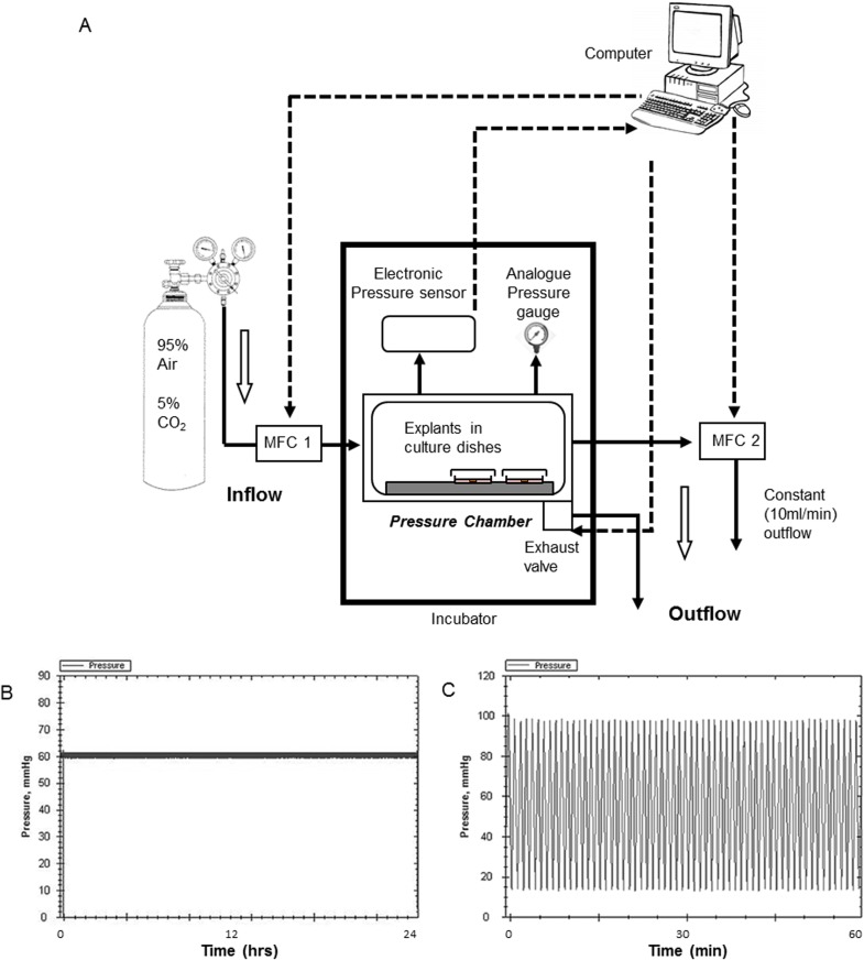
A chamber was designed to expose cells to increased HP (constant and fluctuating). Accurate pressure control (10-100 mmHg) was achieved using mass flow controllers. Human organotypic retinal cultures (HORCs) from donor eyes (<24 h post mortem) were cultured in serum-free DMEM/HamF12. Increased HP was compared to simulated ischemia (oxygen glucose deprivation, OGD). Cell death and apoptosis were measured by LDH and TUNEL assays, RGC marker expression by qRT-PCR (THY-1) and RGC number by immunohistochemistry (NeuN). Activated p38 and JNK were detected by Western blot.
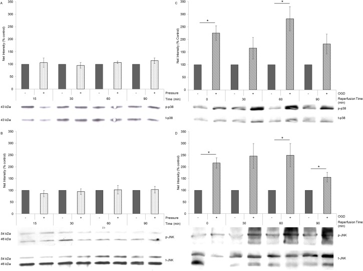
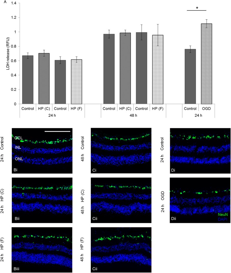
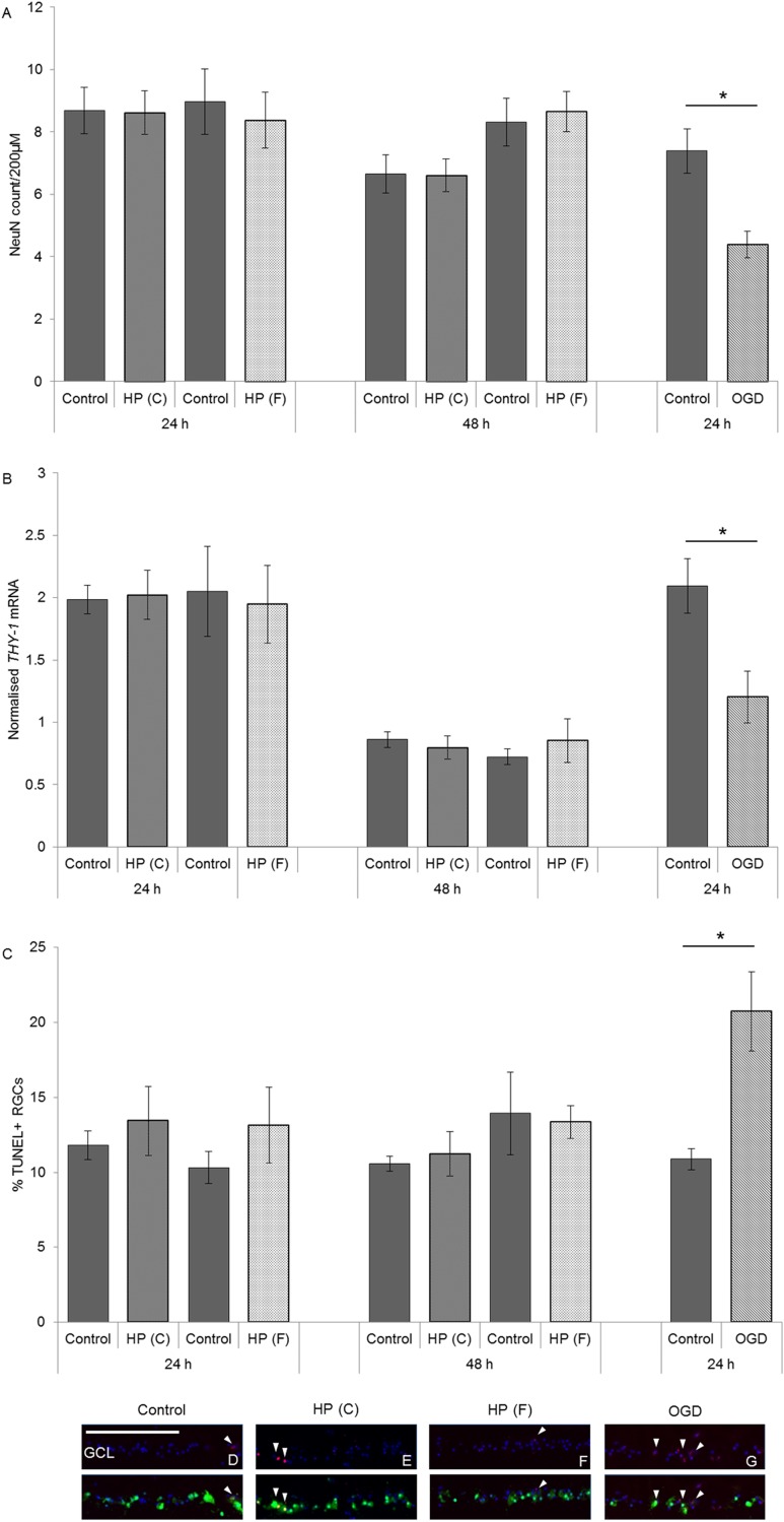
Exposure of HORCs to constant (60 mmHg) or fluctuating (10-100 mmHg; 1 cycle/min) pressure for 24 or 48 h caused no loss of structural integrity, LDH release, decrease in RGC marker expression (THY-1) or loss of RGCs compared with controls. In addition, there was no increase in TUNEL-positive NeuN-labelled cells at either time-point indicating no increase in apoptosis of RGCs. OGD increased apoptosis, reduced RGC marker expression and RGC number and caused elevated LDH release at 24 h. p38 and JNK phosphorylation remained unchanged in HORCs exposed to fluctuating pressure (10-100 mmHg; 1 cycle/min) for 15, 30, 60 and 90 min durations, whereas OGD (3 h) increased activation of p38 and JNK, remaining elevated for 90 min post-OGD.
Directly applied HP had no detectable impact on RGC survival and stress-signalling in HORCs. Simulated ischemia, however, activated stress pathways and caused RGC death. These results show that direct HP does not cause degeneration of RGCs in the ex vivo human retina.
眼压升高是青光眼的主要危险因素。眼压升高的一个后果是眼组织受到更高的静水压力(HP)。本研究探讨了升高的HP对人视网膜应激通路信号传导和视网膜神经节细胞(RGC)存活的影响。
设计一个腔室,使细胞暴露于升高的HP(恒定和波动)下。使用质量流量控制器实现精确的压力控制(10 - 100 mmHg)。将来自供体眼(死后<24小时)的人视网膜组织块培养物(HORCs)培养在无血清的DMEM/HamF12中。将升高的HP与模拟缺血(氧糖剥夺,OGD)进行比较。通过乳酸脱氢酶(LDH)和末端脱氧核苷酸转移酶介导的缺口末端标记(TUNEL)测定法测量细胞死亡和凋亡,通过定量逆转录聚合酶链反应(qRT-PCR)(THY-1)测量RGC标志物表达,通过免疫组织化学(NeuN)测量RGC数量。通过蛋白质印迹法检测活化的p38和应激活化蛋白激酶(JNK)。
与对照组相比,将HORCs暴露于恒定压力(60 mmHg)或波动压力(10 - 100 mmHg;每分钟1个循环)24或48小时,未导致结构完整性丧失、LDH释放、RGC标志物表达(THY-1)降低或RGC丢失。此外,在两个时间点,TUNEL阳性的NeuN标记细胞均未增加,表明RGC凋亡未增加。OGD增加了细胞凋亡,降低了RGC标志物表达和RGC数量,并在24小时时导致LDH释放增加。在暴露于波动压力(10 - 100 mmHg;每分钟1个循环)15、30、60和90分钟的HORCs中,p38和JNK磷酸化保持不变,而OGD(3小时)增加了p38和JNK的活化,在OGD后90分钟仍保持升高。
直接施加HP对HORCs中的RGC存活和应激信号传导没有可检测到的影响。然而,模拟缺血激活了应激通路并导致RGC死亡。这些结果表明,直接HP不会导致离体人视网膜中的RGC退化。