Miklavc Pika, Ehinger Konstantin, Sultan Ayesha, Felder Tatiana, Paul Patrick, Gottschalk Kay-Eberhard, Frick Manfred
Department of General Physiology, University of Ulm, Albert-Einstein Allee 11, 89081 Ulm, Germany.
Institute for Experimental Physics, University of Ulm, Albert-Einstein Allee 11, 89081 Ulm, Germany.
J Cell Sci. 2015 Mar 15;128(6):1193-203. doi: 10.1242/jcs.165571. Epub 2015 Jan 30.
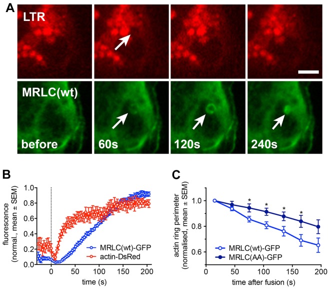
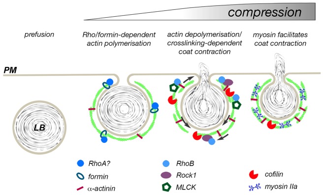
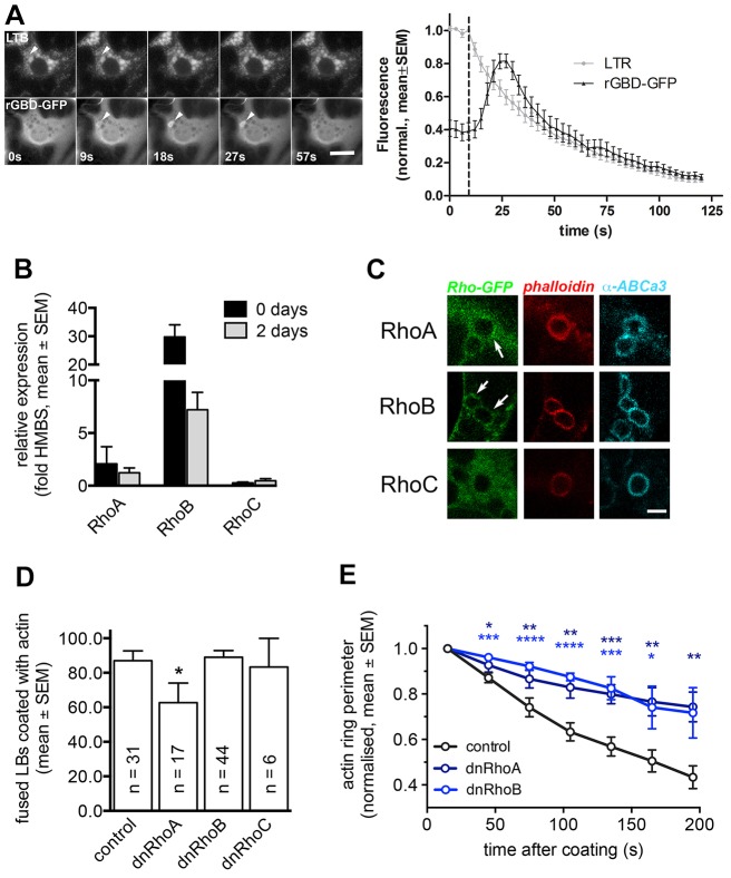
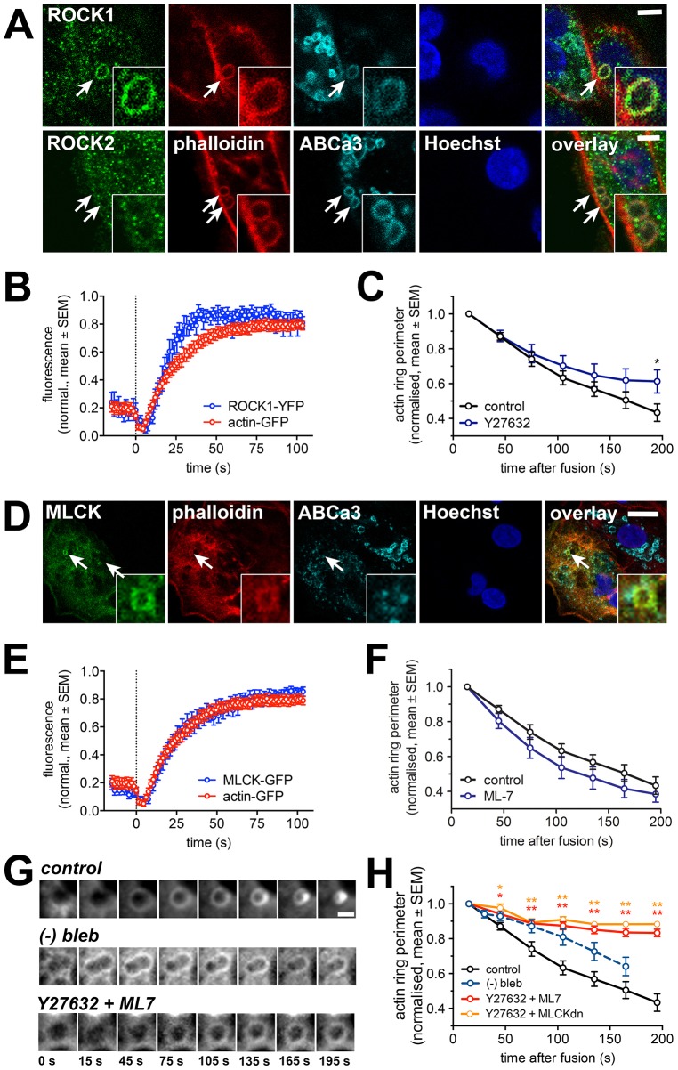
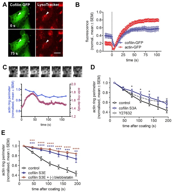
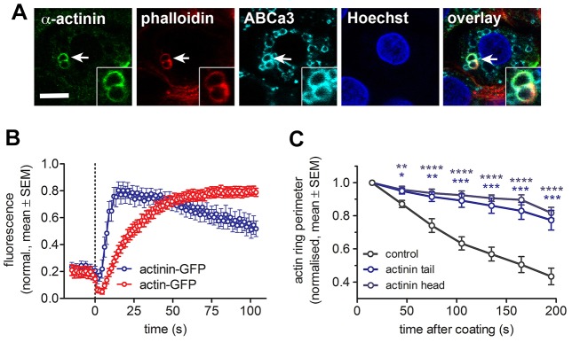
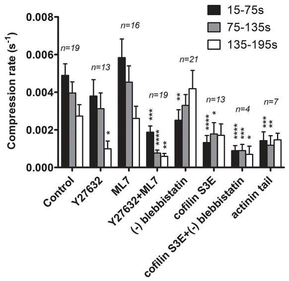
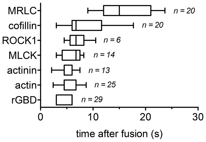
In many secretory cells actin and myosin are specifically recruited to the surface of secretory granules following their fusion with the plasma membrane. Actomyosin-dependent compression of fused granules is essential to promote active extrusion of cargo. However, little is known about molecular mechanisms regulating actin coat formation and contraction. Here, we provide a detailed kinetic analysis of the molecules regulating actin coat contraction on fused lamellar bodies in primary alveolar type II cells. We demonstrate that ROCK1 and myosin light chain kinase 1 (MLCK1, also known as MYLK) translocate to fused lamellar bodies and activate myosin II on actin coats. However, myosin II activity is not sufficient for efficient actin coat contraction. In addition, cofilin-1 and α-actinin translocate to actin coats. ROCK1-dependent regulated actin depolymerisation by cofilin-1 in cooperation with actin crosslinking by α-actinin is essential for complete coat contraction. In summary, our data suggest a complementary role for regulated actin depolymerisation and crosslinking, and myosin II activity, to contract actin coats and drive secretion.
在许多分泌细胞中,肌动蛋白和肌球蛋白在分泌颗粒与质膜融合后会特异性地募集到分泌颗粒表面。肌动球蛋白依赖的融合颗粒压缩对于促进货物的主动挤出至关重要。然而,关于调节肌动蛋白包被形成和收缩的分子机制知之甚少。在这里,我们对调节原代肺泡II型细胞中融合板层小体上肌动蛋白包被收缩的分子进行了详细的动力学分析。我们证明,ROCK1和肌球蛋白轻链激酶1(MLCK1,也称为MYLK)转位到融合板层小体并激活肌动蛋白包被上的肌球蛋白II。然而,肌球蛋白II的活性不足以实现有效的肌动蛋白包被收缩。此外,丝切蛋白-1和α-辅肌动蛋白转位到肌动蛋白包被上。ROCK1依赖的丝切蛋白-1调节的肌动蛋白解聚与α-辅肌动蛋白的肌动蛋白交联协同作用对于完全的包被收缩至关重要。总之,我们的数据表明,调节的肌动蛋白解聚和交联以及肌球蛋白II的活性在收缩肌动蛋白包被和驱动分泌方面具有互补作用。