Shen Yanguang, Sun Zhonghua, Xu Lei, Li Yu, Zhang Nan, Yan Zixu, Fan Zhanming
Department of Radiology, Beijing Anzhen Hospital, Capital Medical University-Beijing Institute of Heart Lung and Blood Vessel Diseases, Beijing, China; Department of Radiology, Hospital Affiliated to Hainan Medical College, Haikou, City of Hainan Province, China.
Discipline of Medical Imaging, Department of Imaging and Applied Physics, Curtin University, Perth, Australia.
PLoS One. 2015 Feb 2;10(2):e0117469. doi: 10.1371/journal.pone.0117469. eCollection 2015.
To assess the image quality of aorta obtained by dual-source computed tomography angiography (DSCTA), performed with high pitch, low tube voltage, and low iodine concentration contrast medium (CM) with images reconstructed using iterative reconstruction (IR).
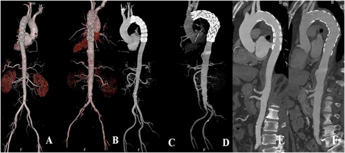
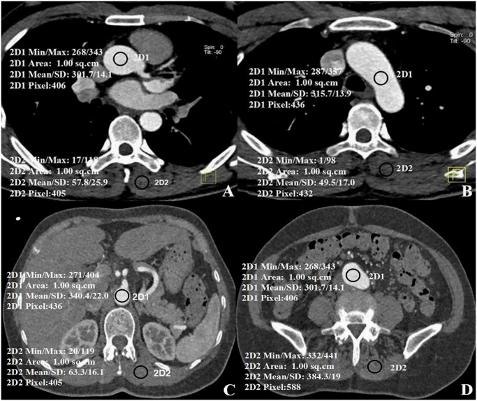
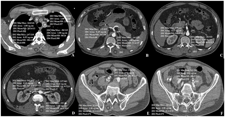
One hundred patients randomly allocated to receive one of two types of CM underwent DSCTA with the electrocardiogram-triggered Flash protocol. In the low-iodine group, 50 patients received CM containing 270 mg I/mL and were scanned at low tube voltage (100 kVp). In the high-iodine CM group, 50 patients received CM containing 370 mg I/mL and were scanned at the tube voltage (120 kVp). The filtered back projection (FBP) algorithm was used for reconstruction in both groups. In addition, the IR algorithm was used in the low-iodine group. Image quality of the aorta was analyzed subjectively by a 3-point grading scale and objectively by measuring the CT attenuation in terms of the signal- and contrast-to-noise ratios (SNR and CNR, respectively). Radiation and CM doses were compared.
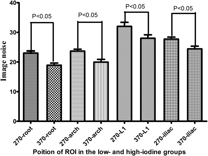
The CT attenuation, subjective image quality assessment, SNR, and CNR of various aortic regions of interest did not differ significantly between two groups. In the low-iodine group, images reconstructed by FBP and IR demonstrated significant differences in image noise, SNR, and CNR (p<0.05). The low-iodine group resulted in 34.3% less radiation (4.4 ± 0.5 mSv) than the high-iodine group (6.7 ± 0.6 mSv), and 27.3% less iodine weight (20.36 ± 2.65 g) than the high-iodine group (28 ± 1.98 g). Observers exhibited excellent agreement on the aortic image quality scores (κ = 0.904).
CT images of aorta could be obtained within 2 s by using a DSCT Flash protocol with low tube voltage, IR, and low-iodine-concentration CM. Appropriate contrast enhancement was achieved while maintaining good image quality and decreasing the radiation and iodine doses.
评估采用高螺距、低管电压及低碘浓度对比剂(CM)的双源计算机断层血管造影(DSCTA)结合迭代重建(IR)技术重建图像时,主动脉的图像质量。
100例患者随机分为两组,分别接受两种类型的CM,采用心电图触发的Flash扫描协议进行DSCTA检查。低碘组50例患者接受含碘量为270 mg I/mL的CM,采用低管电压(100 kVp)扫描。高碘CM组50例患者接受含碘量为370 mg I/mL的CM,采用管电压(120 kVp)扫描。两组均采用滤波反投影(FBP)算法进行重建。此外,低碘组采用IR算法。通过3分制主观评分分析主动脉的图像质量,并通过测量CT衰减值计算信号噪声比(SNR)和对比噪声比(CNR)进行客观分析。比较辐射剂量和CM剂量。
两组不同主动脉感兴趣区的CT衰减值、主观图像质量评估、SNR和CNR差异均无统计学意义。在低碘组,FBP和IR重建的图像在图像噪声、SNR和CNR方面存在显著差异(p<0.05)。低碘组的辐射剂量(4.4±0.5 mSv)比高碘组(6.7±0.6 mSv)减少34.3%,碘用量(20.36±2.65 g)比高碘组(28±1.98 g)减少27.3%。观察者对主动脉图像质量评分的一致性良好(κ = 0.904)。
采用低管电压、IR和低碘浓度CM的DSCT Flash扫描协议,可在2秒内获得主动脉的CT图像。在保持良好图像质量的同时,实现了适当的对比增强,降低了辐射剂量和碘用量。