Feyzi-Dehkhargani Sajad, Shahrooz Rasoul, Malekinejad Hassan, Sadrkhanloo Rajab-Ali
Department of Comparative Histology & Embryology, Faculty of Veterinary Medicine, Urmia University, Urmia, Iran;
Department of Pharmacology and Toxicology, Faculty of Veterinary Medicine, Urmia University, Urmia, Iran.
Vet Res Forum. 2012 Winter;3(1):19-26.
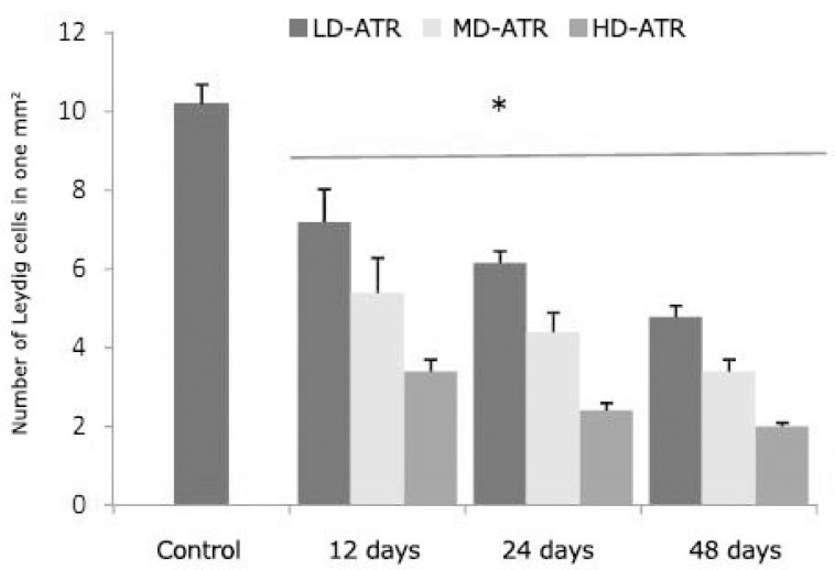
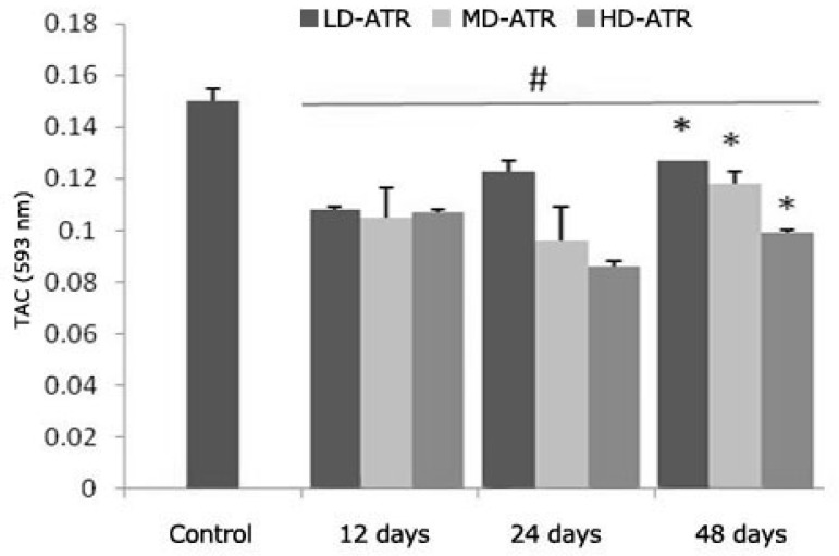
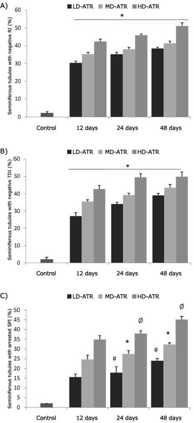
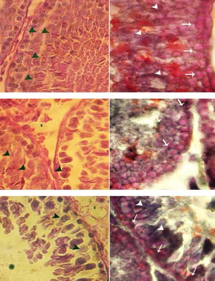
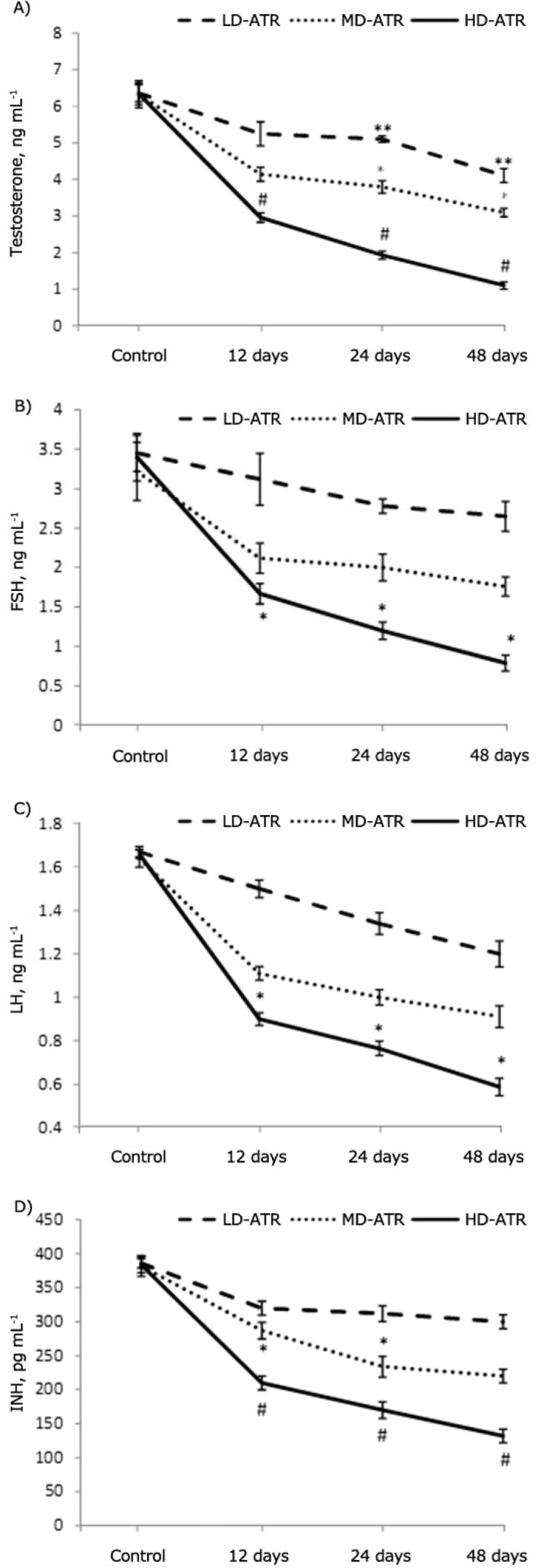
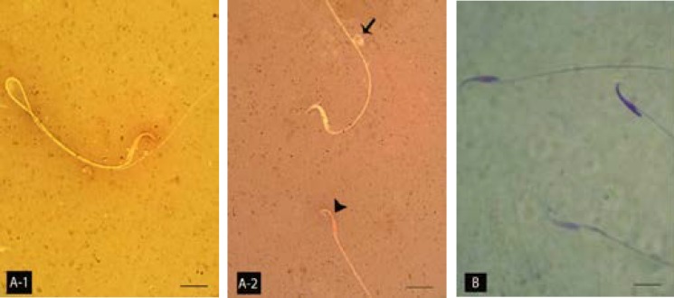
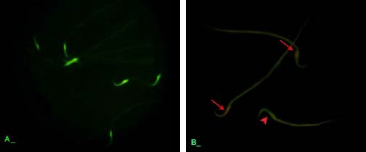
This study was designed to evaluate the detrimental effect of atrazine (ATR) on germinal epitheliums (GE) cytoplasmic carbohydrate (CH) and unsaturated fatty acids (UFA) ratio and to clarify the effect of ATR on serum levels of FSH, LH, testosterone and inhibin-B (INH-B). The impact of ATR exposure on total antioxidant capacity (TAC), sperm DNA packing and integrity were also investigated. Seventy two Wistar rats were used. The rats in control group received vehicle and the animals in test groups received 100, 200 and 300 mg kg(-1) BW of ATR orally on daily bases for 12, 24 and 48 days. In ATR-received groups the spermatogenesis cell were presented with dense reactive sites for lipidophilic staining associated with faint cytoplasmic CH accumulation. Dissociated germinal epithelium, negative tubular and repopulation indexes were manifested. The serum levels of testosterone, FSH, LH and INH-B decreased by 85% after 48 days exposure to high dose of ATR. TAC was reduced in a time- and dose-dependent manner. The sperm DNA damage was marked in animals which exposed to high dose of ATR (72.50 ± 2.25%) and the percentage of nuclear immature sperm increased up to 83.40 ± 0.89%. In conclusion, ATR not only induced its detrimental effect on the endocrine function of the testes and pituitary gland but also affected the cytoplasmic CH ratio and consequently leads to inadequate energy supplement in spermatogenesis cells. Therefore the imbalanced oxidative stress occurs in testicular tissue, which in turn enhances the sperm DNA disintegrity and nuclear immaturity.
本研究旨在评估莠去津(ATR)对生精上皮(GE)细胞质碳水化合物(CH)和不饱和脂肪酸(UFA)比例的有害影响,并阐明ATR对血清促卵泡生成素(FSH)、促黄体生成素(LH)、睾酮和抑制素B(INH - B)水平的影响。还研究了ATR暴露对总抗氧化能力(TAC)、精子DNA包装和完整性的影响。使用了72只Wistar大鼠。对照组大鼠接受赋形剂,试验组动物每天口服100、200和300 mg kg⁻¹体重的ATR,持续12、24和48天。在接受ATR的组中,生精细胞呈现出与微弱细胞质CH积累相关的亲脂性染色密集反应位点。出现了生精上皮解离、阴性管腔和再增殖指数。暴露于高剂量ATR 48天后,睾酮、FSH、LH和INH - B的血清水平下降了85%。TAC以时间和剂量依赖的方式降低。在暴露于高剂量ATR的动物中精子DNA损伤明显(72.50 ± 2.25%),核未成熟精子的百分比增加至83.40 ± 0.89%。总之,ATR不仅对睾丸和垂体的内分泌功能产生有害影响,还影响细胞质CH比例,从而导致生精细胞能量补充不足。因此,睾丸组织中发生氧化应激失衡,进而增强精子DNA完整性破坏和核未成熟。