Fukuda Hiromitsu, Hidaka Teruo, Takagi-Akiba Miyuki, Ichimura Koichiro, Oliva Trejo Juan Alejandro, Sasaki Yu, Wang Juan, Sakai Tatsuo, Asanuma Katsuhiko, Tomino Yasuhiko
Division of Nephrology, Department of Internal Medicine, Juntendo University School of Medicine, 2-1-1, Hongo, Bunkyo-ku, Tokyo, 113-8421, Japan.
Cell Tissue Res. 2015 May;360(2):391-400. doi: 10.1007/s00441-014-2100-9. Epub 2015 Feb 13.
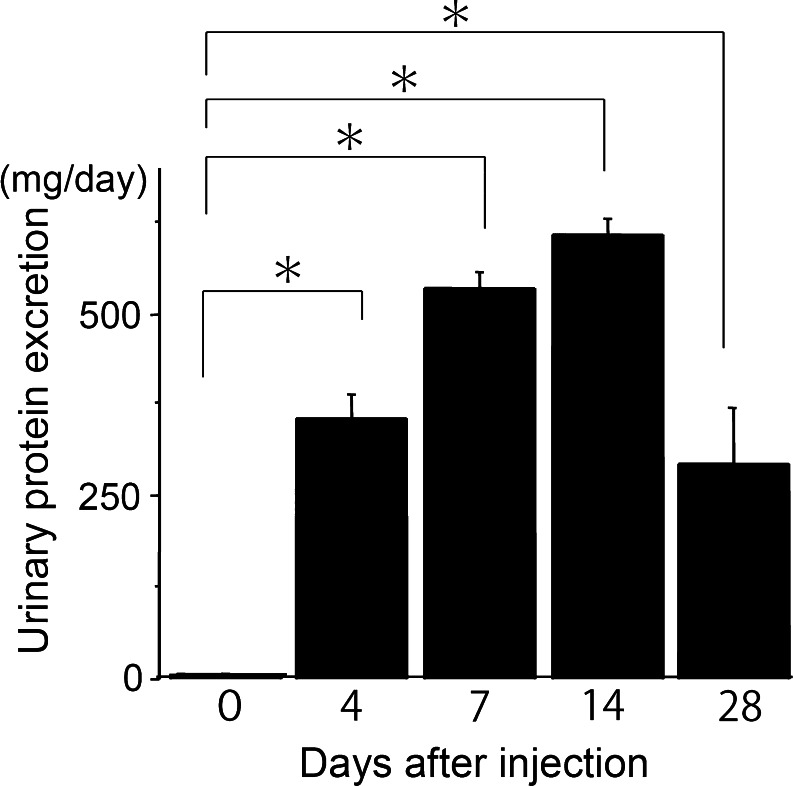
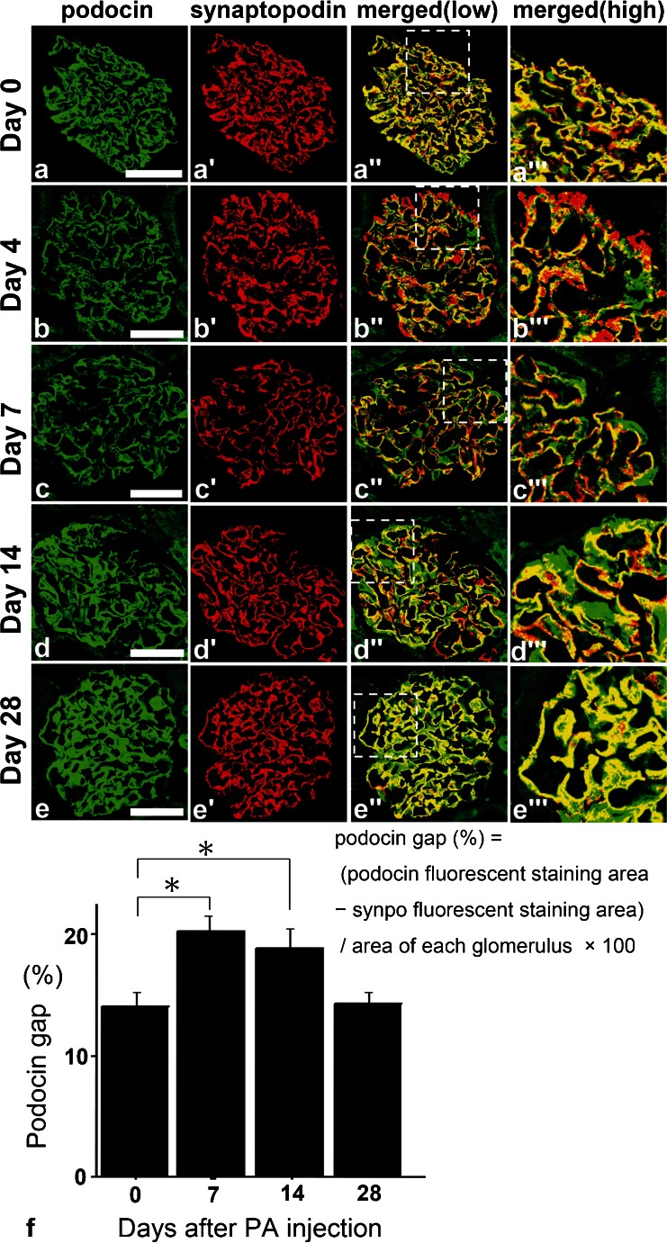
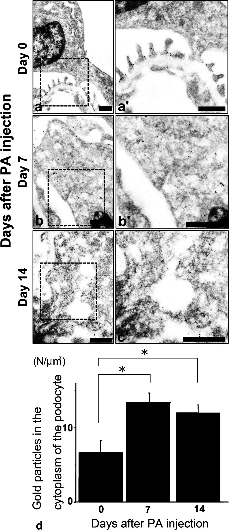
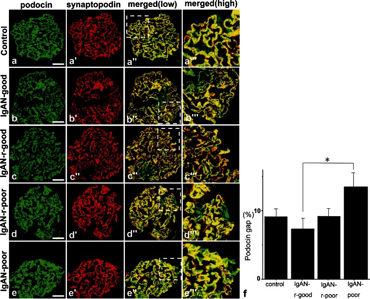
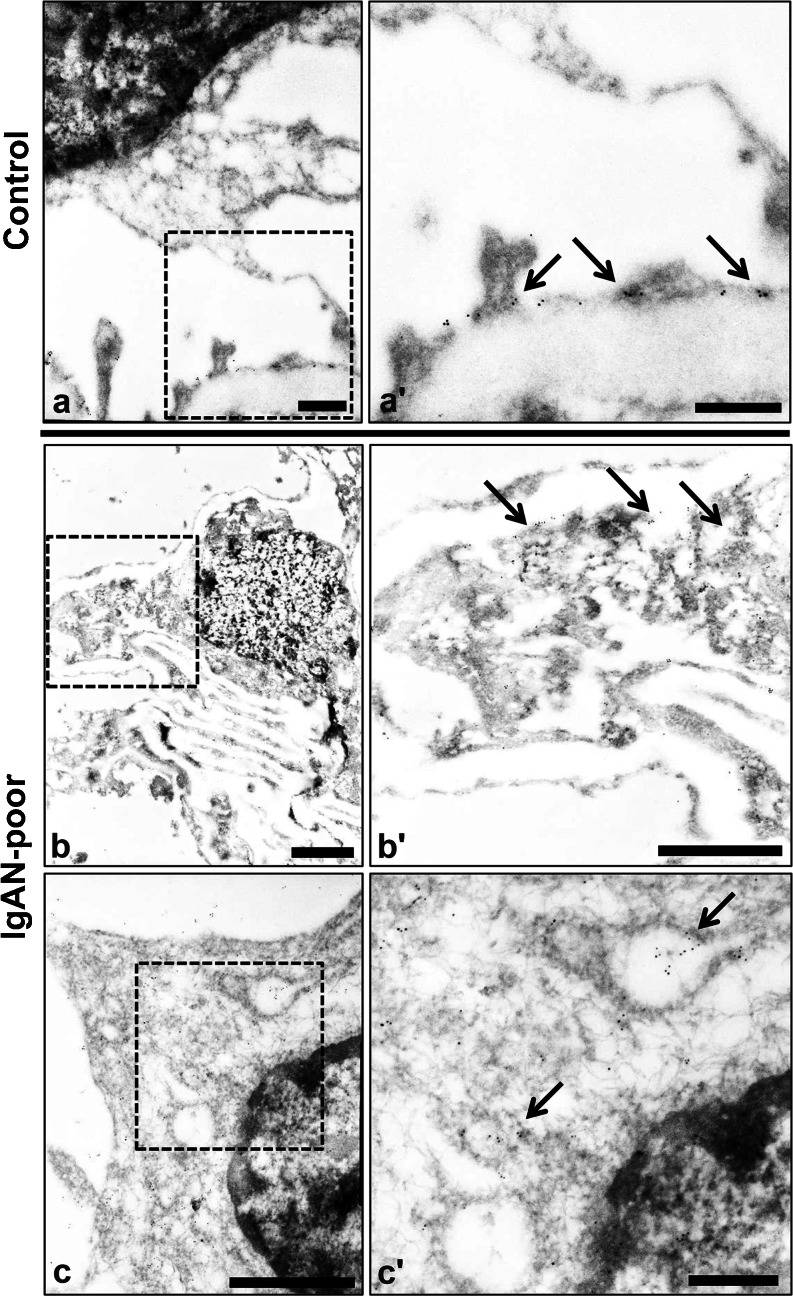
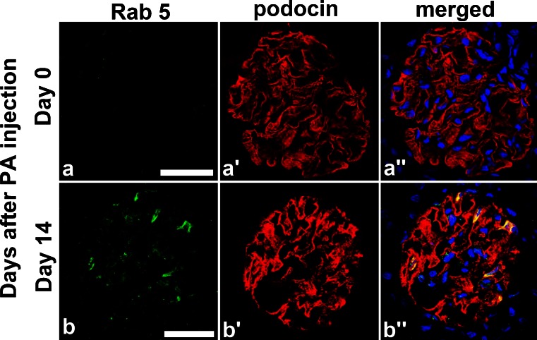
Podocytes serve as the final barrier to urinary protein loss through a highly specialized structure called a slit membrane and maintain foot process and glomerular basement membranes. Podocyte injury results in progressive glomerular damage and accelerates sclerotic changes, although the exact mechanism of podocyte injury is still obscure. We focus on the staining gap (podocin gap) defined as the staining difference between podocin and synaptopodin, which are normally located in the foot process. In puromycin aminonucleoside nephrosis rats, the podocin gap is significantly increased (p < 0.05) and podocin is translocated to the cytoplasm on days 7 and 14 but not on day 28. Surprisingly, the gap is also significantly increased (p < 0.05) in human kidney biopsy specimens of poor-prognosis IgA nephropathy patients. This suggests that the podocin gap could be a useful marker for classifying the prognosis of IgA nephropathy and indicating the translocation of podocin to the cytoplasm. Next, we find more evidence of podocin trafficking in podocytes where podocin merges with Rab5 in puromycin aminonucleoside nephrosis rats at day 14. In immunoelectron microscopy, the podocin positive area was significantly translocated from the foot process areas to the cytoplasm (p< 0.05) on days 7 and 14 in puromycin aminonucleoside nephrosis rats. Interestingly, podocin is also translocated to the cytoplasm in poor-prognosis human IgA nephropathy. In this paper, we demonstrate that the translocation of podocin by endocytosis could be a key traffic event of critical podocyte injury and that the podocin gap could indicate the prognosis of IgA nephropathy.
足细胞通过一种称为裂孔膜的高度特化结构,成为阻止尿蛋白流失的最后一道屏障,并维持足突和肾小球基底膜。尽管足细胞损伤的确切机制仍不清楚,但足细胞损伤会导致肾小球进行性损伤并加速硬化改变。我们关注的是染色间隙(足动蛋白间隙),它被定义为通常位于足突的足动蛋白和突触素之间的染色差异。在嘌呤霉素氨基核苷肾病大鼠中,足动蛋白间隙显著增加(p < 0.05),并且在第7天和第14天足动蛋白转移至细胞质,但在第28天没有。令人惊讶的是,在预后不良的IgA肾病患者的肾活检标本中,该间隙也显著增加(p < 0.05)。这表明足动蛋白间隙可能是用于对IgA肾病预后进行分类以及指示足动蛋白向细胞质转移的有用标志物。接下来,我们发现了更多足细胞中足动蛋白转运的证据,在嘌呤霉素氨基核苷肾病大鼠第14天时,足动蛋白与Rab5融合。在免疫电子显微镜下,嘌呤霉素氨基核苷肾病大鼠在第7天和第14天时,足动蛋白阳性区域从足突区域显著转移至细胞质(p < 0.05)。有趣的是,在预后不良的人类IgA肾病中足动蛋白也转移至细胞质。在本文中,我们证明通过内吞作用导致的足动蛋白转运可能是关键足细胞损伤的关键转运事件,并且足动蛋白间隙可能指示IgA肾病的预后。