Department of Cell Biology, Kidney Research Center, Niigata University Graduate School of Medical and Dental Sciences, 1-757 Asahimachi-dori, Chuo-ku, Niigata, 951-8510, Japan.
Clin Exp Nephrol. 2020 Mar;24(3):193-204. doi: 10.1007/s10157-020-01854-3. Epub 2020 Feb 4.
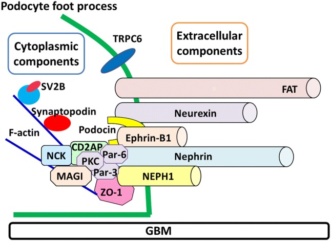
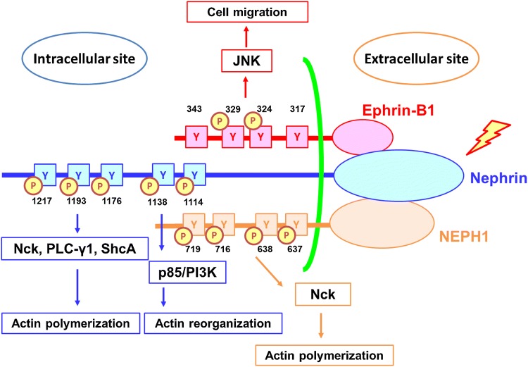
Dysfunction of slit diaphragm, a cell-cell junction of glomerular podocytes, is involved in the development of proteinuria in several glomerular diseases. Slit diaphragm should be a target of a novel therapy for proteinuria. Nephrin, NEPH1, P-cadherin, FAT, and ephrin-B1 were reported to be extracellular components forming a molecular sieve of the slit diaphragm. Several cytoplasmic proteins such as ZO-1, podocin, CD2AP, MAGI proteins and Par-complex molecules were identified as scaffold proteins linking the slit diaphragm to the cytoskeleton. In this article, new insights into these molecules and the pathogenic roles of the dysfunction of these molecules were introduced. The slit diaphragm functions not only as a barrier but also as a signaling platform transfer the signal to the inside of the cell. For maintaining the slit diaphragm function properly, the phosphorylation level of nephrin is strictly regulated. The recent studies on the signaling pathway from nephrin, NEPH1, and ephrin-B1 were reviewed. Although the mechanism regulating the function of the slit diaphragm had remained unclear, recent studies revealed TRPC6 and angiotensin II-regulating mechanisms play a critical role in regulating the barrier function of the slit diaphragm. In this review, recent investigations on the regulation of the slit diaphragm function were reviewed, and a strategy for the establishment of a novel therapy for proteinuria was proposed.
裂孔隔膜功能障碍,肾小球足细胞的细胞-细胞连接,参与几种肾小球疾病蛋白尿的发生。裂孔隔膜应该是蛋白尿新型治疗的靶点。研究报道,nephrin、NEPH1、P-钙黏蛋白、FAT 和 ephrin-B1 是形成裂孔隔膜分子筛的细胞外成分。几种细胞质蛋白,如 ZO-1、podocin、CD2AP、MAGI 蛋白和 Par 复合物分子,被鉴定为将裂孔隔膜与细胞骨架连接的支架蛋白。在本文中,介绍了这些分子的新见解,以及这些分子功能障碍的致病作用。裂孔隔膜不仅作为屏障,而且作为信号转导平台,将信号传递到细胞内部。为了保持裂孔隔膜的正常功能,nephrin 的磷酸化水平受到严格调节。本文综述了关于 nephrin、NEPH1 和 ephrin-B1 的信号通路的最新研究。尽管调节裂孔隔膜功能的机制尚不清楚,但最近的研究揭示了 TRPC6 和血管紧张素 II 调节机制在调节裂孔隔膜的屏障功能中起着关键作用。在这篇综述中,回顾了最近对裂孔隔膜功能调节的研究,并提出了一种建立蛋白尿新型治疗策略的设想。