Gotecha Sarang, Ranade Deepak, Sharma Shrikant, Punia Prashant, Kotecha Megha
Department of Neurosurgery, Padmashree Dr. D. Y. Patil Hospital, Pimpri, Pune, Maharashtra, India.
Asian J Neurosurg. 2014 Oct-Dec;9(4):244. doi: 10.4103/1793-5482.146653.
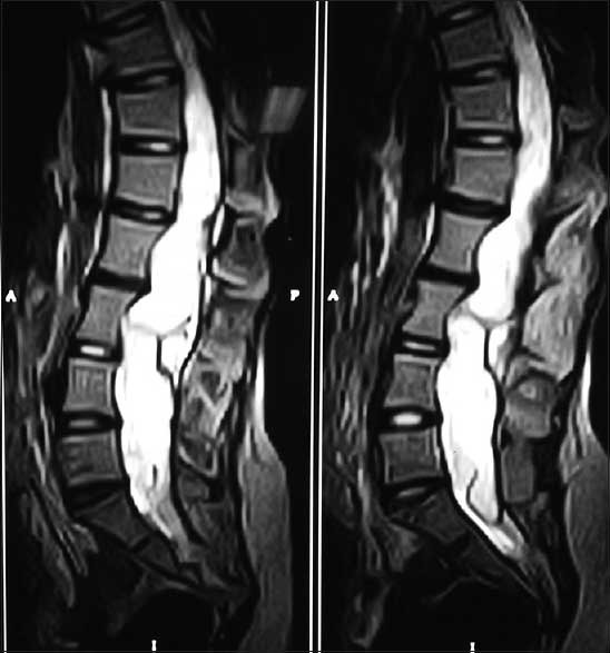
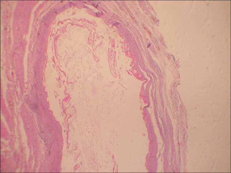
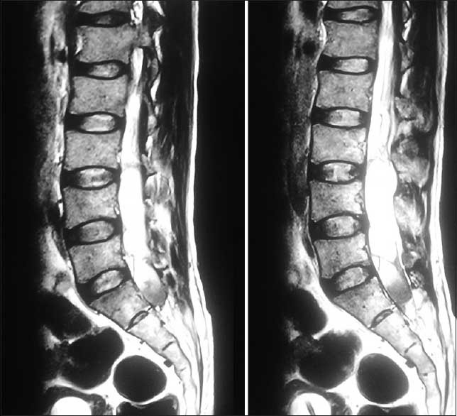
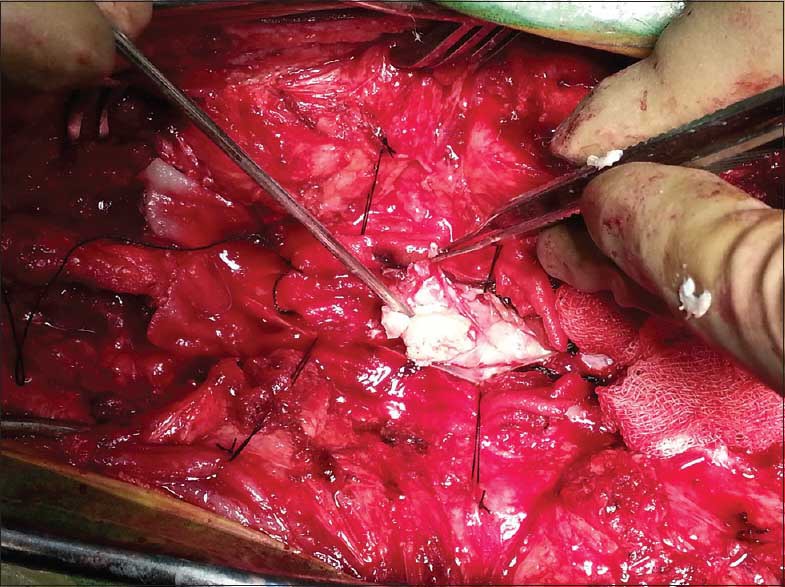
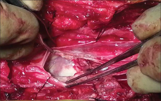
We report two cases with giant intramedullary epidermoid cysts in the thoracolumbosacral and lumbosacral regions with varied presentations. Magnetic resonance (MR) imaging of the thoraco lumbar spine in case 1revealed an intramedullary elongated mass extending from T10 to S2 level causing significant widening of the spinal canal while MR imaging of lumbosacral spine in case 2 showed straightening of the lumbar spine and spina bifida at L5 level with conus at L3 and a lobulated long segment intramedullary solid cystic lesion extending from L2 to S2 veterbrae. The lesion was surgically resected and the pathology revealed an epidermoid cyst. Epidermoid cysts of the spinal cord are rare tumours in the adult population which may be congenital or acquired. Symptoms arising from epidermoid cysts vary with the level of involvement. The treatment of epidermoid cysts is surgical and if possible, complete removal is the goal.
我们报告了两例分别位于胸腰段和腰骶段的巨大髓内表皮样囊肿病例,其表现各异。病例1胸腰椎的磁共振成像显示,髓内有一细长肿物,从T10延伸至S2水平,导致椎管显著增宽;而病例2腰骶椎的磁共振成像显示腰椎变直,L5水平有脊柱裂,圆锥位于L3,还有一个分叶状长节段髓内实性囊性病变,从L2延伸至S2椎体。病变经手术切除,病理显示为表皮样囊肿。脊髓表皮样囊肿在成人中是罕见肿瘤,可能是先天性或后天性的。表皮样囊肿引起的症状因受累部位而异。表皮样囊肿的治疗方法是手术,若可能,目标是完整切除。