Kehoe Elizabeth G, Farrell Dervla, Metzler-Baddeley Claudia, Lawlor Brian A, Kenny Rose Anne, Lyons Declan, McNulty Jonathan P, Mullins Paul G, Coyle Damien, Bokde Arun L
Trinity College Institute of Neuroscience and Cognitive Systems Group, Discipline of Psychiatry, School of Medicine, Trinity College Dublin , Dublin , Ireland.
Cardiff University Brain Research Imaging Centre (CUBRIC), Neuroscience and Mental Health Research Institute (NMHRI), School of Psychology, Cardiff University , Cardiff , UK.
Front Aging Neurosci. 2015 Feb 5;7:10. doi: 10.3389/fnagi.2015.00010. eCollection 2015.
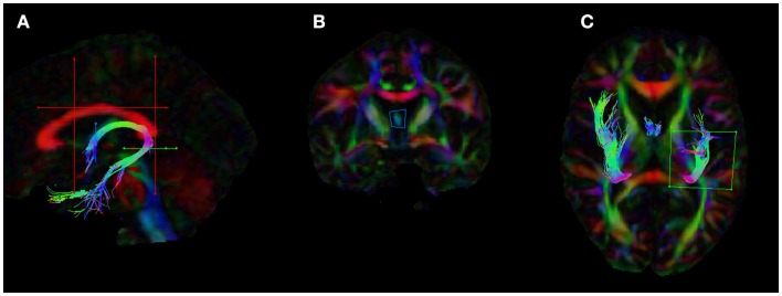
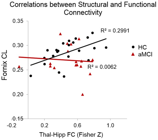
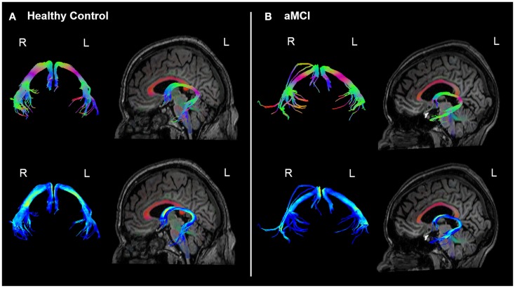
In this study, we wished to examine the relationship between the structural connectivity of the fornix, a white matter (WM) tract in the limbic system, which is affected in amnestic mild cognitive impairment (aMCI) and Alzheimer's disease, and the resting-state functional connectivity (FC) of two key related subcortical structures, the thalamus, and hippocampus. Twenty-two older healthy controls (HC) and 18 older adults with aMCI underwent multi-modal MRI scanning. The fornix was reconstructed using constrained-spherical deconvolution-based tractography. The FC between the thalamus and hippocampus was calculated using a region-of-interest approach from which the mean time series were exacted and correlated. Diffusion tensor imaging measures of the WM microstructure of the fornix were correlated against the Fisher Z correlation values from the FC analysis. There was no difference between the groups in the fornix WM measures, nor in the resting-state FC of the thalamus and hippocampus. We did however find that the relationship between functional and structural connectivity differed significantly between the groups. In the HCs, there was a significant positive association between linear diffusion (CL) in the fornix and the FC of the thalamus and hippocampus, however, there was no relationship between these measures in the aMCI group. These preliminary findings suggest that in aMCI, the relationship between the functional and structural connectivity of regions of the limbic system may be significantly altered compared to healthy ageing. The combined use of diffusion weighted imaging and functional MRI may advance our understanding of neural network changes in aMCI, and elucidate subtle changes in the relationship between structural and functional brain networks.
在本研究中,我们希望考察穹窿(边缘系统中的一个白质束,在遗忘型轻度认知障碍(aMCI)和阿尔茨海默病中会受到影响)的结构连通性与两个关键相关皮质下结构——丘脑和海马体的静息态功能连通性(FC)之间的关系。22名健康老年人(HC)和18名患有aMCI的老年人接受了多模态磁共振成像扫描。使用基于约束球面反卷积的纤维束成像重建穹窿。使用感兴趣区域方法计算丘脑和海马体之间的FC,从中提取平均时间序列并进行相关性分析。穹窿白质微结构的扩散张量成像测量值与FC分析中的费舍尔Z相关值进行相关性分析。两组在穹窿白质测量方面以及丘脑和海马体的静息态FC方面均无差异。然而,我们确实发现两组之间功能连通性和结构连通性的关系存在显著差异。在健康对照组中,穹窿中的线性扩散(CL)与丘脑和海马体的FC之间存在显著正相关,然而,在aMCI组中这些测量值之间没有关系。这些初步发现表明,在aMCI中,与健康衰老相比,边缘系统区域的功能连通性和结构连通性之间的关系可能会发生显著改变。扩散加权成像和功能磁共振成像的联合使用可能会增进我们对aMCI中神经网络变化的理解,并阐明脑结构网络和功能网络之间关系的细微变化。