Reccia Isabella, Pisanu Adolfo, Podda Mauro, Uccheddu Alessandro
From the Department of Surgery, Clinica Chirurgica, University of Cagliari, Policlinico Universitario di Monserrato, Sestu (CA), Italy (IR, AP, MP, AU).
Medicine (Baltimore). 2015 Feb;94(7):e319. doi: 10.1097/MD.0000000000000319.
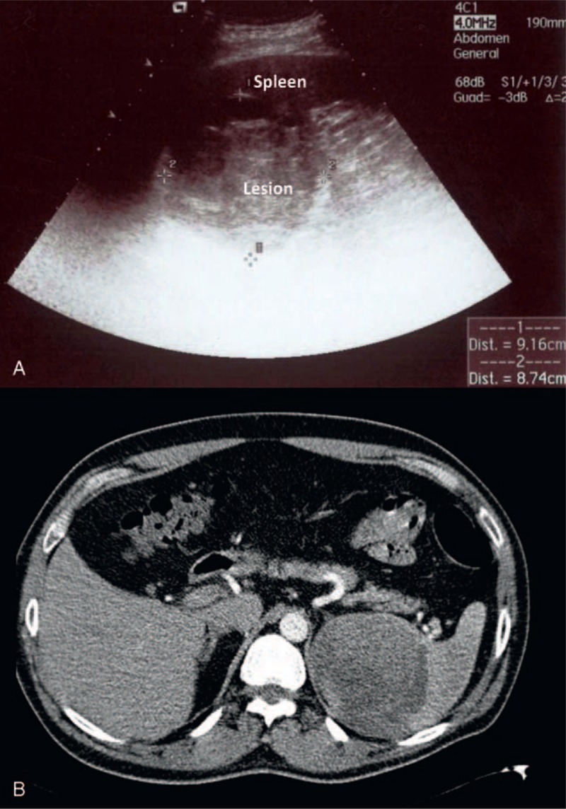
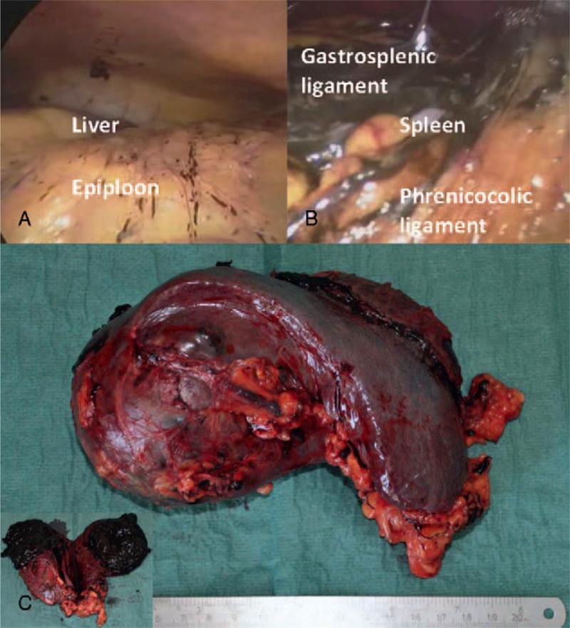
Metastases to the spleen are rare and are generally part of a multi-visceral metastatic disease. The most common sources of splenic metastases include breast, lung and colorectal malignancies as well as melanoma and ovarian carcinoma. Solitary splenic metastasis is very uncommon. We present a case of a 44-year-old man who presented at our department for gallstones symptoms. He had a past medical history of neck cutaneous melanoma (T3bN0M0--Stage IIb). He had not attended follow-up schedule for personal reasons. However, abdominal ultrasound revealed the presence of a solitary solid lesion in the spleen. Preoperative workup was completed with CT scan that confirmed the presence of a large splenic lesion with subcapsular fluid collection, also compatible with a post-traumatic lesion.Preoperative findings could not exclude malignancy and patient was therefore submitted to surgery. At laparoscopy, a condition of peritoneal melanosis was present. Splenectomy was carried out. Histological report confirmed the peritoneal melanosis and the diagnosis of metastatic spleen lesion from melanoma. Patient was observed, but died of metastatic disease 14 months after surgery. Splenic metastases are uncommon. Isolated metastases from melanoma are rare and could be found several months after primary diagnosis of melanoma. Surgery remains the most effective treatment, especially for metachronous disease, offering the best chance of long-term survival. Prognosis remains poor, as metachronous disease is indicative of aggressive widespread of the disease.
脾脏转移瘤罕见,通常是多脏器转移性疾病的一部分。脾脏转移瘤最常见的来源包括乳腺癌、肺癌和结直肠癌,以及黑色素瘤和卵巢癌。孤立性脾脏转移非常少见。我们报告一例44岁男性患者,因胆结石症状前来我院就诊。他既往有颈部皮肤黑色素瘤病史(T3bN0M0——IIb期)。因个人原因他未按随访计划就诊。然而,腹部超声显示脾脏存在一个孤立性实性病变。术前通过CT扫描完成了检查,CT扫描证实存在一个大的脾脏病变,伴有包膜下液体积聚,也符合创伤后病变。术前检查结果不能排除恶性肿瘤,因此患者接受了手术。在腹腔镜检查时,发现存在腹膜黑变病。进行了脾切除术。组织学报告证实了腹膜黑变病以及黑色素瘤脾脏转移病变的诊断。对患者进行了观察,但患者在手术后14个月死于转移性疾病。脾脏转移瘤并不常见。黑色素瘤的孤立转移罕见,可在黑色素瘤初次诊断数月后发现。手术仍然是最有效的治疗方法,尤其是对于异时性疾病,提供了长期生存的最佳机会。预后仍然很差,因为异时性疾病表明疾病具有侵袭性的广泛扩散。