Wollborn Jakob, Wunder Christian, Stix Jana, Neuhaus Winfried, Bruno Rapahel R, Baar Wolfgang, Flemming Sven, Roewer Norbert, Schlegel Nicolas, Schick Martin A
Department of Anaesthesia and Critical Care, University Hospital Würzburg, Germany.
Department of Pathology, Klinikum Nürnberg, Nürnberg, Germany.
J Pharmacol Pharmacother. 2015 Jan-Mar;6(1):13-23. doi: 10.4103/0976-500X.149138.
To investigate the impact of the phophodiesterase-4 inhibition (PD-4-I) with rolipram on hepatic integrity in lipopolysaccharide (LPS) induced hyperinflammation.
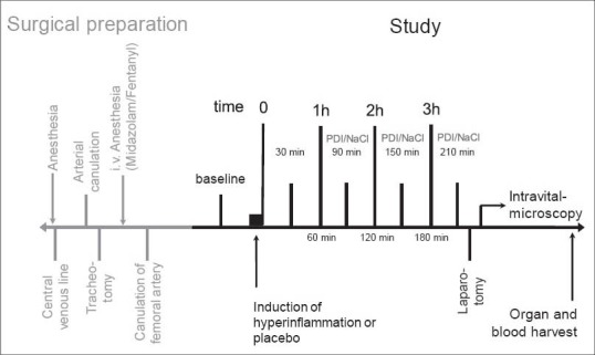
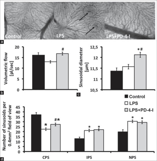
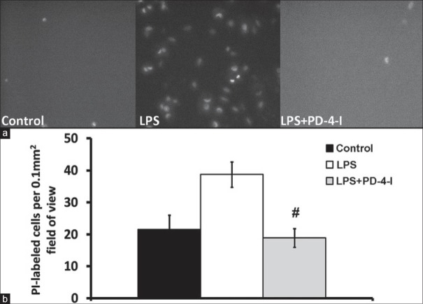
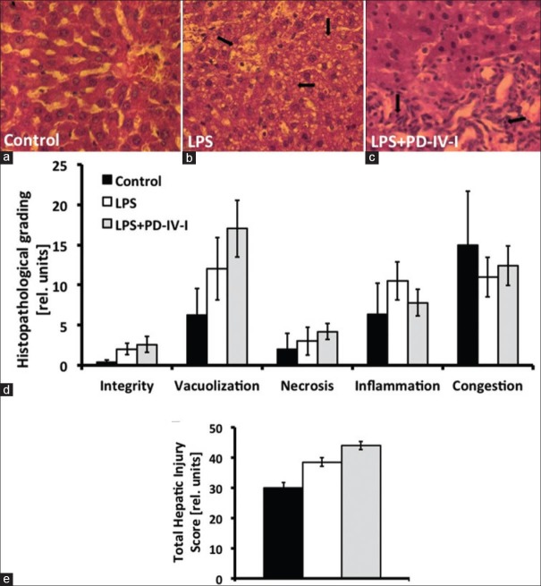
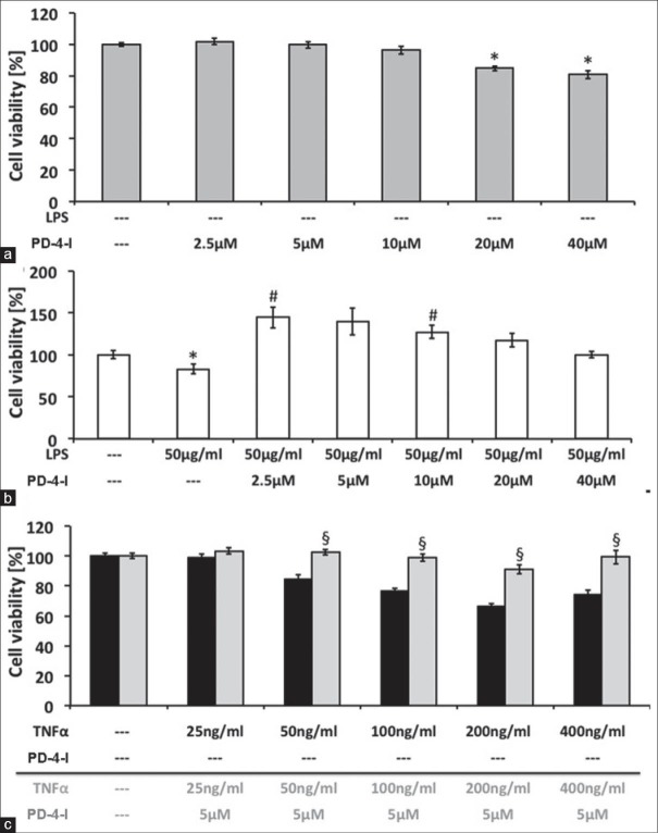
Liver microcirculation in rats was obtained using intravital microscopy. Macrohemodynamic parameters, blood assays, and organs were harvested to determine organ function and injury. Hyperinflammation was induced by LPS and PD-4-I rolipram was administered intravenously one hour after LPS application. Cell viability of HepG2 cells was measured by EZ4U-kit based on the dye XTT. Experiments were carried out assessing the influence of different concentrations of tumor necrosis factor alpha (TNF-α) and LPS with or without PD-4-I.
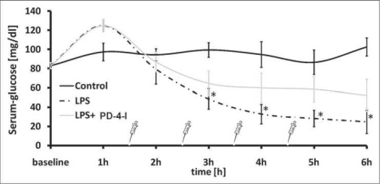
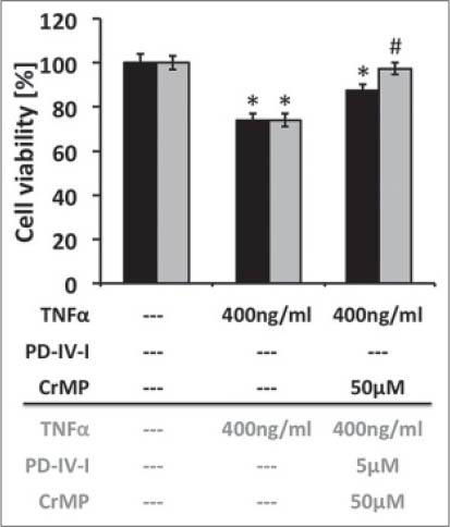
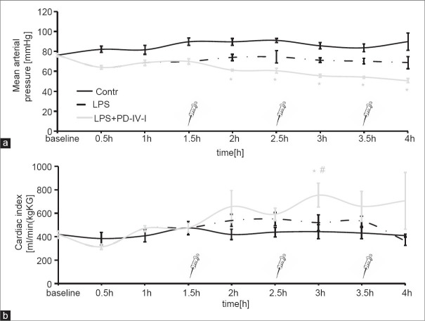
Untreated LPS-induced rats showed significantly decreased liver microcirculation and increased hepatic cell death, whereas LPS + PD-4-I treatment could improve hepatic volumetric flow and cell death to control level whithout influencing the inflammatory impact. In HepG2 cells TNF-α and LPS significantly reduced cell viability. Coincubation with PD-4-I increased HepG2 viability to control levels. The heme oxygenase 1 (HO-1) pathway did not induce the protective effect of PD-4-I.
Intravenous PD-4-I treatment was effective in improving hepatic microcirculation and hepatic integrity, while it had a direct protective effect on HepG2 viability during inflammation.
研究咯利普兰对磷酸二酯酶-4的抑制作用(PD-4-I)对脂多糖(LPS)诱导的肝脏超炎症反应中肝脏完整性的影响。
采用活体显微镜观察大鼠肝脏微循环。采集大鼠的宏观血流动力学参数、血液样本及器官,以确定器官功能和损伤情况。通过注射LPS诱导超炎症反应,并在注射LPS 1小时后静脉注射PD-4-I咯利普兰。基于XTT染料,使用EZ4U试剂盒测定HepG2细胞的活力。开展实验评估不同浓度的肿瘤坏死因子α(TNF-α)和LPS在有或无PD-4-I情况下的影响。
未经处理的LPS诱导大鼠肝脏微循环显著减少,肝细胞死亡增加,而LPS + PD-4-I治疗可将肝脏容积流量和细胞死亡改善至对照水平,且不影响炎症反应。在HepG2细胞中,TNF-α和LPS显著降低细胞活力。与PD-4-I共同孵育可使HepG2细胞活力提高至对照水平。血红素加氧酶1(HO-1)途径未诱导PD-4-I的保护作用。
静脉注射PD-4-I治疗可有效改善肝脏微循环和肝脏完整性,同时在炎症期间对HepG2细胞活力具有直接保护作用。