• 文献检索
  • 文档翻译
  • 深度研究
  • 学术资讯
  • Suppr Zotero 插件Zotero 插件
  • 邀请有礼
  • 套餐&价格
  • 历史记录
应用&插件
Suppr Zotero 插件Zotero 插件浏览器插件Mac 客户端Windows 客户端微信小程序
定价
高级版会员购买积分包购买API积分包
服务
文献检索文档翻译深度研究API 文档MCP 服务
关于我们
关于 Suppr公司介绍联系我们用户协议隐私条款
关注我们

Suppr 超能文献

核心技术专利:CN118964589B侵权必究
粤ICP备2023148730 号-1Suppr @ 2026

文献检索

告别复杂PubMed语法,用中文像聊天一样搜索,搜遍4000万医学文献。AI智能推荐,让科研检索更轻松。

立即免费搜索

文件翻译

保留排版,准确专业,支持PDF/Word/PPT等文件格式,支持 12+语言互译。

免费翻译文档

深度研究

AI帮你快速写综述,25分钟生成高质量综述,智能提取关键信息,辅助科研写作。

立即免费体验

通过磁共振显微镜以10微米各向同性分辨率对果蝇大脑中的突触域进行可视化。

Visualization of synaptic domains in the Drosophila brain by magnetic resonance microscopy at 10 micron isotropic resolution.

作者信息

Lee Choong H, Blackband Stephen J, Fernandez-Funez Pedro

机构信息

Department of Neuroscience, McKnight Brain Institute, University of Florida, Gainesville, FL. 32611, USA.

1] Department of Neuroscience, McKnight Brain Institute, University of Florida, Gainesville, FL. 32611, USA [2] National High Magnetic Field Laboratory, University of Florida, Gainesville, FL. 32611, USA.

出版信息

Sci Rep. 2015 Mar 10;5:8920. doi: 10.1038/srep08920.

DOI:10.1038/srep08920
PMID:25753480
原文链接:https://pmc.ncbi.nlm.nih.gov/articles/PMC4649768/
Abstract

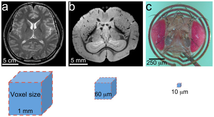
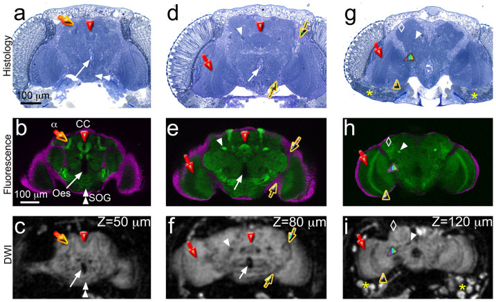
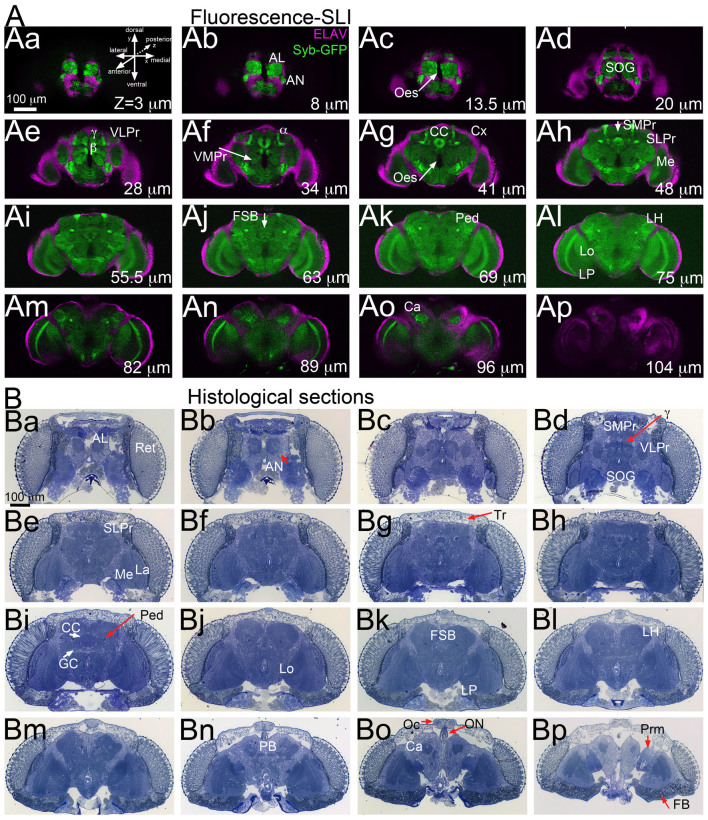
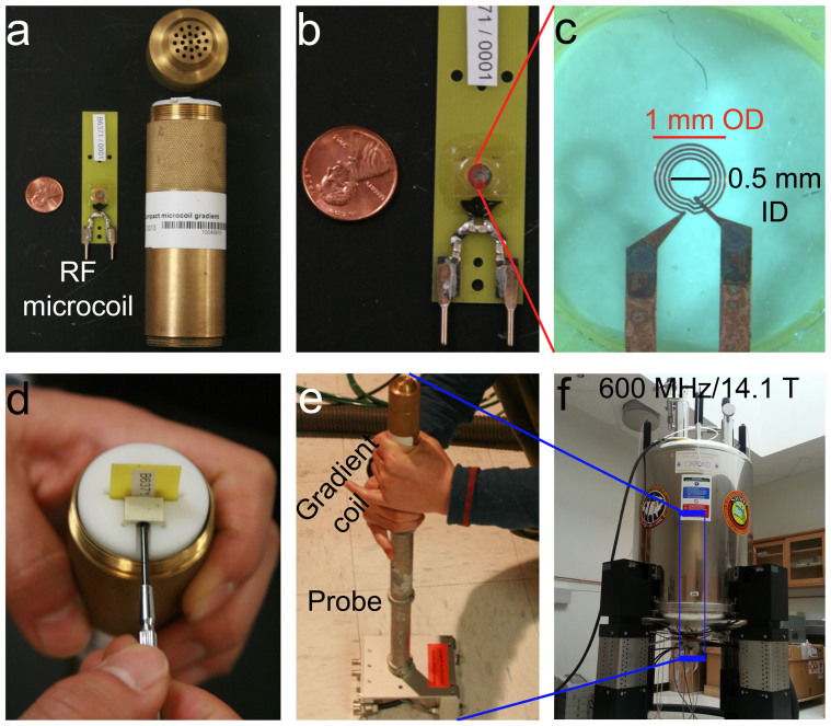
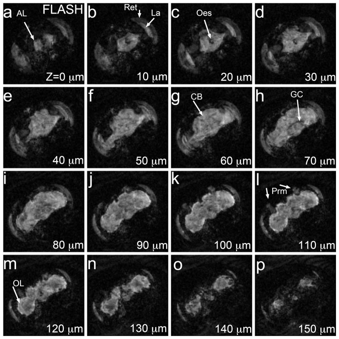
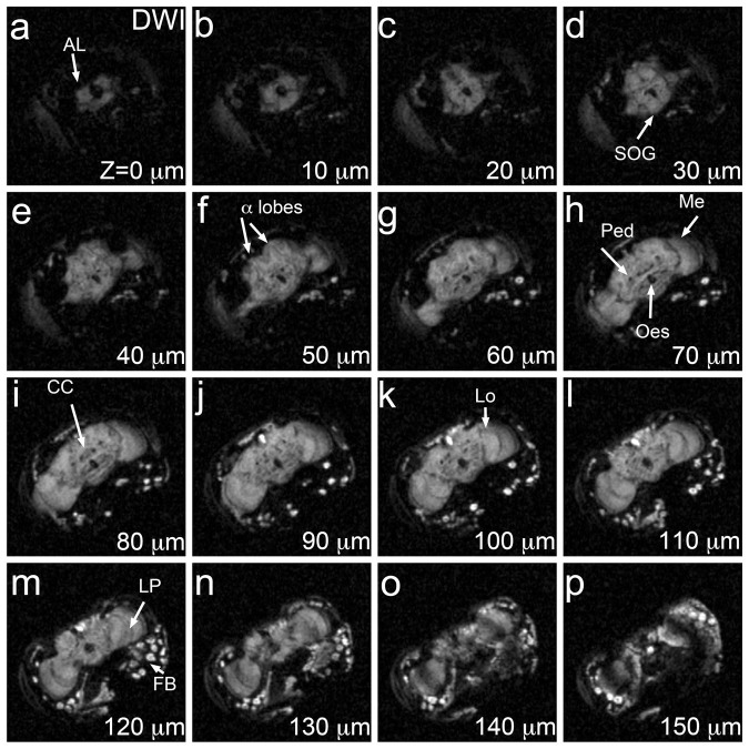
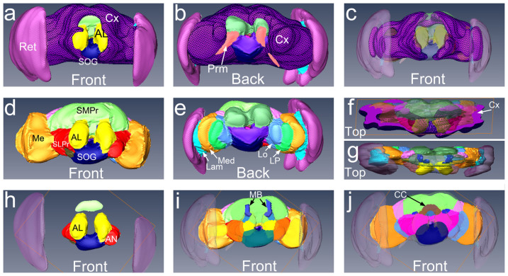
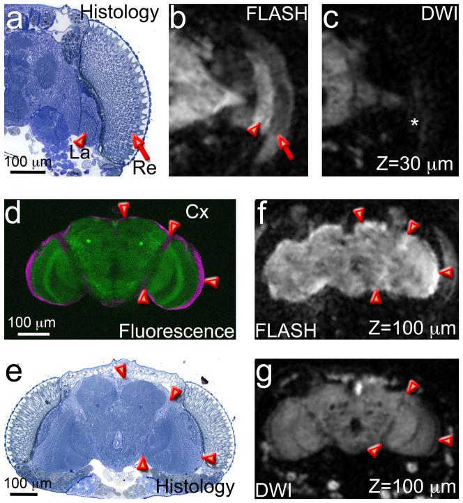
Understanding the complex architecture, connectivity, and pathology of the human brain is a major application of magnetic resonance imaging (MRI). However, the cellular basis of MR signal is still poorly understood. The advent of MR microscopy (MRM) enables imaging biological samples at cellular resolution, helping to interpret the nature of MR signal at the cellular level. In this regard, the small Drosophila brain can reveal key aspects of MR signal through the visualization of complex, intact neuronal structures in their native spatial arrangement. Applying state-of-the-art MR technology, we imaged fixed Drosophila heads at 10 μm isotropic resolution by two endogenously contrasted MRM sequences. The improved MRM sensitivity described here delivered the highest 3D resolution of an intact animal head reported so far. 3D fast low angle shot (FLASH) revealed strong signal in most internal tissues, particularly in the brain cortex, which contains the cell bodies of neurons and glia. Remarkably, 3D diffusion weighted imaging (DWI) delivered unprecedented contrast within the modular brain neuropil, revealing hyperintense signal in synapse-rich microdomains. Thus, the complex Drosophila brain revealed unknown features of FLASH and DWI with potential applications in characterizing the structure and pathology of the mammalian brain.

摘要

了解人类大脑的复杂结构、连接性和病理学是磁共振成像(MRI)的一项主要应用。然而,人们对磁共振信号的细胞基础仍知之甚少。磁共振显微镜(MRM)的出现使得以细胞分辨率对生物样本进行成像成为可能,有助于在细胞水平上解释磁共振信号的本质。在这方面,果蝇的小脑袋能够通过可视化其天然空间排列中的复杂完整神经元结构,揭示磁共振信号的关键方面。应用最先进的磁共振技术,我们通过两个内源性对比的MRM序列,以各向同性10μm的分辨率对固定的果蝇头部进行成像。本文所述的改进后的MRM灵敏度提供了迄今为止报道的完整动物头部的最高三维分辨率。三维快速低角度激发(FLASH)在大多数内部组织中显示出强信号,特别是在包含神经元和神经胶质细胞体的大脑皮层中。值得注意的是,三维扩散加权成像(DWI)在模块化脑髓质内提供了前所未有的对比度,在富含突触的微域中显示出高强度信号。因此,复杂的果蝇大脑揭示了FLASH和DWI的未知特征,在表征哺乳动物大脑的结构和病理学方面具有潜在应用。

https://cdn.ncbi.nlm.nih.gov/pmc/blobs/ea3e/4649768/2ca4998eb7c5/srep08920-f8.jpg
https://cdn.ncbi.nlm.nih.gov/pmc/blobs/ea3e/4649768/0fd371e4347a/srep08920-f1.jpg
https://cdn.ncbi.nlm.nih.gov/pmc/blobs/ea3e/4649768/107109ee809d/srep08920-f2.jpg
https://cdn.ncbi.nlm.nih.gov/pmc/blobs/ea3e/4649768/954dde9150ef/srep08920-f3.jpg
https://cdn.ncbi.nlm.nih.gov/pmc/blobs/ea3e/4649768/b6811cf7720c/srep08920-f4.jpg
https://cdn.ncbi.nlm.nih.gov/pmc/blobs/ea3e/4649768/09b9ba5fe466/srep08920-f5.jpg
https://cdn.ncbi.nlm.nih.gov/pmc/blobs/ea3e/4649768/d16c6a1e78fd/srep08920-f6.jpg
https://cdn.ncbi.nlm.nih.gov/pmc/blobs/ea3e/4649768/3419b6c16329/srep08920-f7.jpg
https://cdn.ncbi.nlm.nih.gov/pmc/blobs/ea3e/4649768/2ca4998eb7c5/srep08920-f8.jpg

相似文献

1
Visualization of synaptic domains in the Drosophila brain by magnetic resonance microscopy at 10 micron isotropic resolution.通过磁共振显微镜以10微米各向同性分辨率对果蝇大脑中的突触域进行可视化。
Sci Rep. 2015 Mar 10;5:8920. doi: 10.1038/srep08920.
2
Investigation of the subcellular architecture of L7 neurons of Aplysia californica using magnetic resonance microscopy (MRM) at 7.8 microns.使用7.8微米磁共振显微镜(MRM)对加州海兔L7神经元的亚细胞结构进行研究。
Sci Rep. 2015 Jun 10;5:11147. doi: 10.1038/srep11147.
3
Visualization of mouse barrel cortex using ex-vivo track density imaging.使用离体轨迹密度成像技术对小鼠大脑皮层 barrels 区进行可视化。
Neuroimage. 2014 Feb 15;87:465-75. doi: 10.1016/j.neuroimage.2013.09.030. Epub 2013 Sep 21.
4
Magnetic resonance microscopy of human and porcine neurons and cellular processes.磁共振显微镜在人类和猪神经元及细胞过程中的应用。
Neuroimage. 2012 Apr 2;60(2):1404-11. doi: 10.1016/j.neuroimage.2012.01.050. Epub 2012 Jan 14.
5
Black-blood multicontrast imaging of carotid arteries with DANTE-prepared 2D and 3D MR imaging.颈动脉 DANTE 准备 2D 和 3D MR 成像的黑血多对比度成像。
Radiology. 2014 Nov;273(2):560-9. doi: 10.1148/radiol.14131717. Epub 2014 Jun 11.
6
Visualization of live, mammalian neurons during Kainate-infusion using magnetic resonance microscopy.使用磁共振显微镜观察在海人酸输注过程中活的哺乳动物神经元的可视化。
Neuroimage. 2020 Oct 1;219:116997. doi: 10.1016/j.neuroimage.2020.116997. Epub 2020 May 31.
7
Magnetic resonance microscopy of mammalian neurons.哺乳动物神经元的磁共振显微镜检查。
Neuroimage. 2009 Jul 15;46(4):1037-40. doi: 10.1016/j.neuroimage.2009.03.009. Epub 2009 Mar 12.
8
High-resolution cartilage imaging of the knee at 3T: basic evaluation of modern isotropic 3D MR-sequences.3T 膝关节高分辨率软骨成像:现代各向同性 3D MR 序列的基本评估。
Eur J Radiol. 2011 Jun;78(3):398-405. doi: 10.1016/j.ejrad.2010.01.008. Epub 2010 Feb 6.
9
Three-dimensional magnetic resonance observation of cartilage repair tissue (MOCART) score assessed with an isotropic three-dimensional true fast imaging with steady-state precession sequence at 3.0 Tesla.在3.0特斯拉磁场下,采用各向同性三维稳态进动快速成像序列评估软骨修复组织的三维磁共振观察(MOCART)评分。
Invest Radiol. 2009 Sep;44(9):603-12. doi: 10.1097/RLI.0b013e3181b5333c.
10
Mathematical models for the diffusion magnetic resonance signal abnormality in patients with prion diseases.朊病毒病患者扩散磁共振信号异常的数学模型。
Neuroimage Clin. 2014 Nov 29;7:142-54. doi: 10.1016/j.nicl.2014.11.017. eCollection 2015.

引用本文的文献

1
Considerations and recommendations from the ISMRM diffusion study group for preclinical diffusion MRI: Part 2-Ex vivo imaging: Added value and acquisition.国际磁共振医学学会(ISMRM)扩散研究小组关于临床前扩散磁共振成像的考量与建议:第2部分——离体成像:附加价值与采集
Magn Reson Med. 2025 Jun;93(6):2535-2560. doi: 10.1002/mrm.30435. Epub 2025 Mar 4.
2
LYNSU: automated 3D neuropil segmentation of fluorescent images for brains.LYNSU:用于大脑荧光图像的自动化3D神经纤维网分割
Front Neuroinform. 2024 Jul 29;18:1429670. doi: 10.3389/fninf.2024.1429670. eCollection 2024.
3
Micron-scale magnetic resonance imaging based on low temperatures and dynamic nuclear polarization.

本文引用的文献

1
Seeing the forest tree by tree: super-resolution light microscopy meets the neurosciences.见树又见林:超分辨率荧光显微镜技术与神经科学的交汇。
Nat Neurosci. 2013 Jul;16(7):790-7. doi: 10.1038/nn.3403. Epub 2013 Jun 25.
2
Imaging morphogenesis: technological advances and biological insights.成像形态发生:技术进步与生物学见解。
Science. 2013 Jun 7;340(6137):1234168. doi: 10.1126/science.1234168.
3
A GAL4-driver line resource for Drosophila neurobiology.用于果蝇神经生物学的 GAL4 驱动子线资源。
基于低温和动态核极化的微米级磁共振成像。
Prog Nucl Magn Reson Spectrosc. 2023 Nov-Dec;138-139:136-149. doi: 10.1016/j.pnmrs.2023.10.001. Epub 2023 Oct 17.
4
Experimental demonstration of diffusion limitations on resolution and SNR in MR microscopy.MR 显微镜中扩散限制对分辨率和 SNR 的实验论证。
J Magn Reson. 2023 Jul;352:107479. doi: 10.1016/j.jmr.2023.107479. Epub 2023 May 26.
5
An optimized workflow for microCT imaging of formalin-fixed and paraffin-embedded (FFPE) early equine embryos.优化福尔马林固定石蜡包埋(FFPE)早期马胚胎 microCT 成像的工作流程。
Anat Histol Embryol. 2022 Sep;51(5):611-623. doi: 10.1111/ahe.12834. Epub 2022 Jul 18.
6
Advanced high resolution three-dimensional imaging to visualize the cerebral neurovascular network in stroke.高级高分辨率三维成像技术,可用于可视化脑卒中患者的脑神经血管网络。
Int J Biol Sci. 2022 Jan 1;18(2):552-571. doi: 10.7150/ijbs.64373. eCollection 2022.
7
Elucidating the complex organization of neural micro-domains in the locust Schistocerca gregaria using dMRI.使用扩散磁共振成像技术阐明蝗虫 Schistocerca gregaria 中神经微域的复杂结构。
Sci Rep. 2021 Feb 9;11(1):3418. doi: 10.1038/s41598-021-82187-3.
8
three-dimensional magnetic resonance imaging of rat knee osteoarthritis model induced using meniscal transection.使用半月板横断术诱导的大鼠膝骨关节炎模型的三维磁共振成像
J Orthop Translat. 2015 Jun 23;3(3):134-141. doi: 10.1016/j.jot.2015.06.002. eCollection 2015 Jul.
9
Probing the brain with molecular fMRI.分子 fMRI 探测大脑。
Curr Opin Neurobiol. 2018 Jun;50:201-210. doi: 10.1016/j.conb.2018.03.009. Epub 2018 Apr 9.
10
Low-temperature magnetic resonance imaging with 2.8 μm isotropic resolution.具有2.8微米各向同性分辨率的低温磁共振成像。
J Magn Reson. 2018 Feb;287:47-55. doi: 10.1016/j.jmr.2017.12.016. Epub 2017 Dec 20.
Cell Rep. 2012 Oct 25;2(4):991-1001. doi: 10.1016/j.celrep.2012.09.011. Epub 2012 Oct 11.
4
In vivo magnetic resonance microscopy of Drosophilae at 9.4 T.在 9.4T 下对果蝇进行活体磁共振显微镜成像。
Magn Reson Imaging. 2013 Jan;31(1):109-19. doi: 10.1016/j.mri.2012.06.019. Epub 2012 Aug 13.
5
Magnetic resonance microscopy of human and porcine neurons and cellular processes.磁共振显微镜在人类和猪神经元及细胞过程中的应用。
Neuroimage. 2012 Apr 2;60(2):1404-11. doi: 10.1016/j.neuroimage.2012.01.050. Epub 2012 Jan 14.
6
Three-dimensional reconstruction of brain-wide wiring networks in Drosophila at single-cell resolution.以单细胞分辨率重建果蝇全脑连接网络的三维结构。
Curr Biol. 2011 Jan 11;21(1):1-11. doi: 10.1016/j.cub.2010.11.056. Epub 2010 Dec 2.
7
Cellular-level diffusion tensor microscopy and fiber tracking in mammalian nervous tissue with direct histological correlation.哺乳动物神经组织的细胞水平扩散张量显微镜和纤维示踪与直接组织学相关性。
Neuroimage. 2010 Aug 15;52(2):556-61. doi: 10.1016/j.neuroimage.2010.04.031. Epub 2010 Apr 18.
8
100 years of Drosophila research and its impact on vertebrate neuroscience: a history lesson for the future.100 年的果蝇研究及其对脊椎动物神经科学的影响:为未来提供的历史教训。
Nat Rev Neurosci. 2010 Jul;11(7):514-22. doi: 10.1038/nrn2839.
9
Genetic dissection of the mouse CNS using magnetic resonance microscopy.利用磁共振显微镜对小鼠中枢神经系统进行基因剖析。
Curr Opin Neurol. 2009 Aug;22(4):379-86. doi: 10.1097/WCO.0b013e32832d9b86.
10
Twin-spot MARCM to reveal the developmental origin and identity of neurons.双斑MARCM技术揭示神经元的发育起源和特性。
Nat Neurosci. 2009 Jul;12(7):947-53. doi: 10.1038/nn.2345. Epub 2009 Jun 14.