Eloot Liesbeth, Thierens Hubert, Taeymans Yves, Drieghe Benny, De Pooter Jan, Van Peteghem Sylvie, Buytaert Dimitri, Gijs Thomas, Lapere Régine, Bacher Klaus
Department of Basic Medical Sciences, Ghent University, Ghent, Belgium.
Heart Centre, Ghent University Hospital, Ghent, Belgium.
Catheter Cardiovasc Interv. 2015 Nov;86(5):E205-12. doi: 10.1002/ccd.25913. Epub 2015 Mar 30.
The purpose of this study was to quantify the reduction in patient radiation dose during coronary angiography (CA) by a new X-ray technology, and to assess its impact on diagnostic image quality.
Recently, a novel X-ray imaging technology has become available for interventional cardiology, using advanced image processing and an optimized acquisition chain for radiation dose reduction.
70 adult patients were randomly assigned to a reference X-ray system or the novel X-ray system. Patient demographics were registered and exposure parameters were recorded for each radiation event. Clinical image quality was assessed for both patient groups.
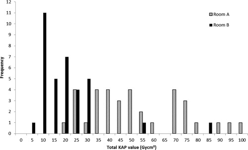
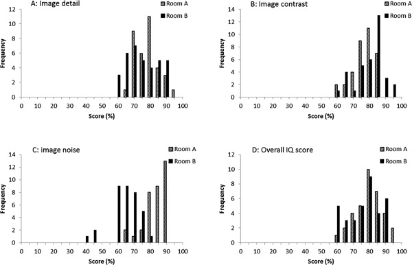
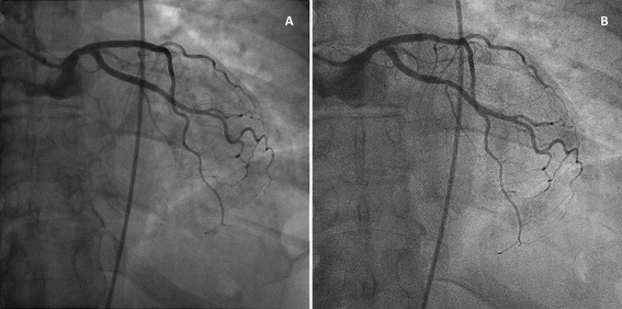
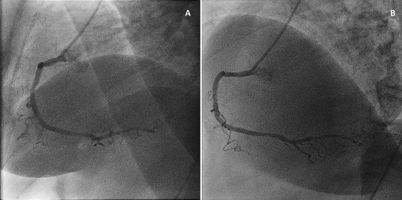
With the same angiographic technique and a comparable patient population, the new imaging technology was associated with a 75% reduction in total kerma-area product (KAP) value (decrease from 47 Gycm2 to 12 Gycm2, P<0.001). Clinical image quality showed an equivalent detail and contrast for both imaging systems. On the other hand, the subjective appreciation of noise was more apparent in images of the new image processing system, acquired at lower doses, compared to the reference system. However, the higher noise content did not affect the overall image quality score, which was adequate for diagnosis in both systems.
For the first time, we present a new X-ray imaging technology, combining advanced noise reduction algorithms and an optimized acquisition chain, which reduces patient radiation dose in CA drastically (75%), while maintaining diagnostic image quality. Use of this technology may further improve the radiation safety of cardiac angiography and interventions.
本研究旨在量化一种新型X射线技术在冠状动脉造影(CA)过程中患者辐射剂量的降低情况,并评估其对诊断图像质量的影响。
最近,一种新型X射线成像技术已应用于介入心脏病学领域,该技术采用先进的图像处理和优化的采集链来降低辐射剂量。
70名成年患者被随机分配至参考X射线系统或新型X射线系统。记录患者人口统计学数据,并记录每次辐射事件的曝光参数。对两组患者的临床图像质量进行评估。
在相同的血管造影技术和相当的患者群体中,新的成像技术使总比释动能面积乘积(KAP)值降低了75%(从47 Gycm²降至12 Gycm²,P<0.001)。两种成像系统的临床图像质量在细节和对比度方面相当。另一方面,与参考系统相比,在较低剂量下采集的新图像处理系统图像中,噪声的主观感受更明显。然而,较高的噪声含量并未影响整体图像质量评分,两个系统的图像质量均足以用于诊断。
我们首次展示了一种新型X射线成像技术,该技术结合了先进的降噪算法和优化的采集链,在冠状动脉造影中可大幅降低患者辐射剂量(75%),同时保持诊断图像质量。使用该技术可能会进一步提高心脏血管造影和介入治疗的辐射安全性。