Andronic Joseph, Shirakashi Ryo, Pickel Simone U, Westerling Katherine M, Klein Teresa, Holm Thorge, Sauer Markus, Sukhorukov Vladimir L
Department of Biotechnology and Biophysics, University of Würzburg, Biozentrum, Am Hubland, Würzburg, Germany.
Institute of Industrial Science, The University of Tokyo, Tokyo, Japan.
PLoS One. 2015 Mar 10;10(3):e0119990. doi: 10.1371/journal.pone.0119990. eCollection 2015.
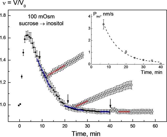
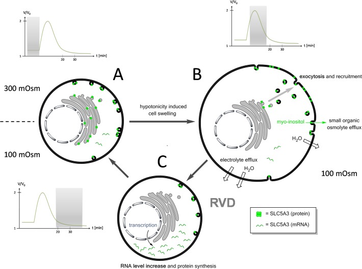
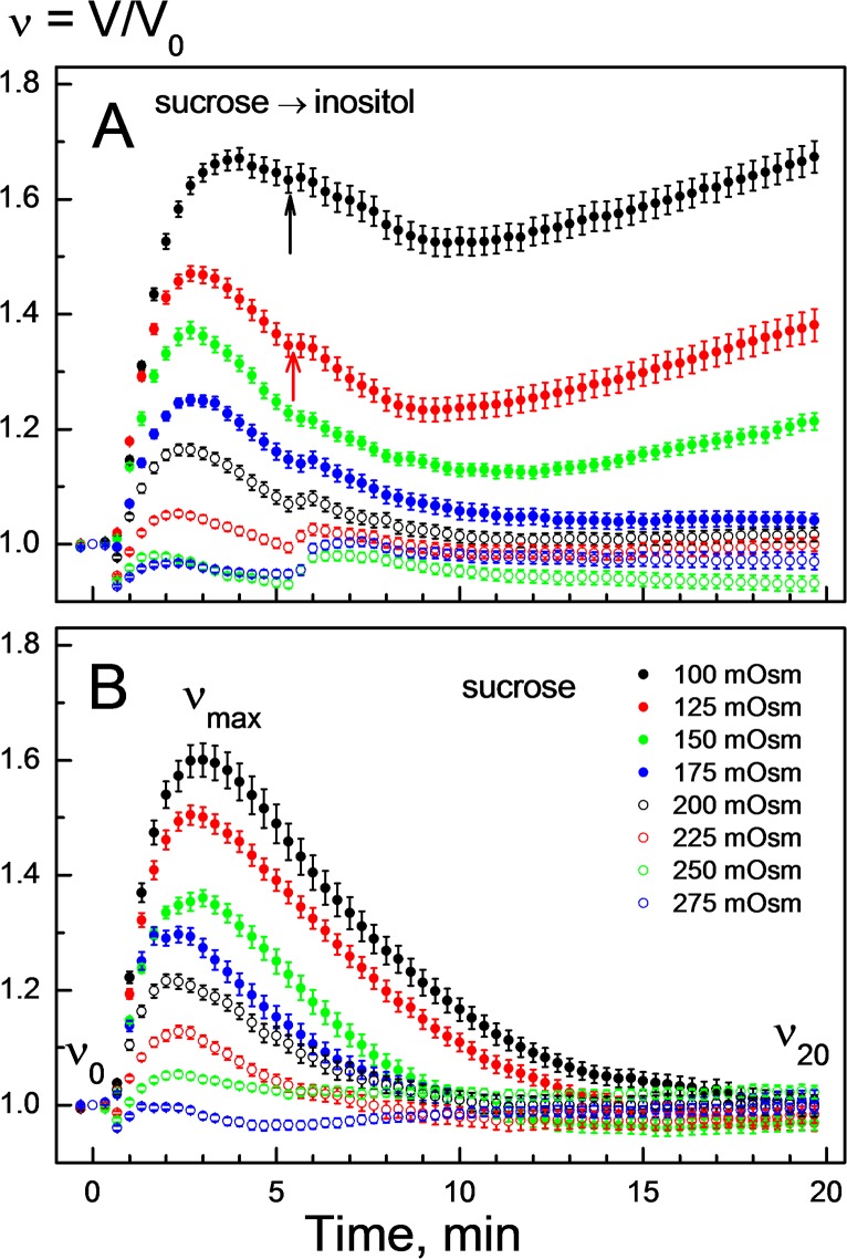
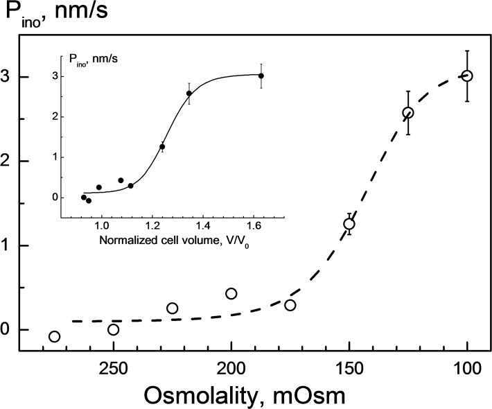
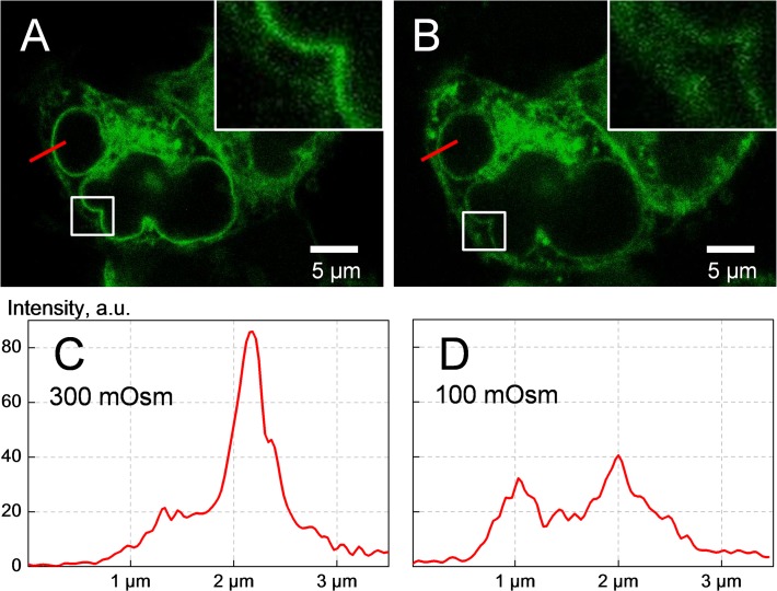
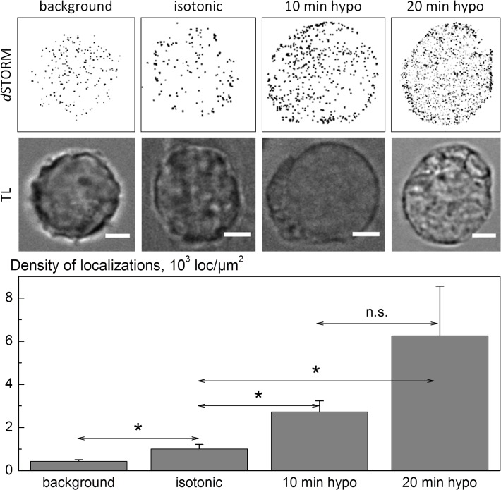
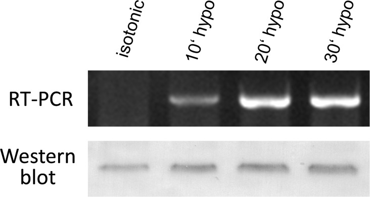
Swelling-activated pathways for myo-inositol, one of the most abundant organic osmolytes in mammalian cells, have not yet been identified. The present study explores the SLC5A3 protein as a possible transporter of myo-inositol in hyponically swollen HEK293 cells. To address this issue, we examined the relationship between the hypotonicity-induced changes in plasma membrane permeability to myo-inositol P ino [m/s] and expression/localization of SLC5A3. P ino values were determined by cell volumetry over a wide tonicity range (100-275 mOsm) in myo-inositol-substituted solutions. While being negligible under mild hypotonicity (200-275 mOsm), P ino grew rapidly at osmolalities below 200 mOsm to reach a maximum of ∼ 3 nm/s at 100-125 mOsm, as indicated by fast cell swelling due to myo-inositol influx. The increase in P ino resulted most likely from the hypotonicity-mediated incorporation of cytosolic SLC5A3 into the plasma membrane, as revealed by confocal fluorescence microscopy of cells expressing EGFP-tagged SLC5A3 and super-resolution imaging of immunostained SLC5A3 by direct stochastic optical reconstruction microscopy (dSTORM). dSTORM in hypotonic cells revealed a surface density of membrane-associated SLC5A3 proteins of 200-2000 localizations/μm2. Assuming SLC5A3 to be the major path for myo-inositol, a turnover rate of 80-800 myo-inositol molecules per second for a single transporter protein was estimated from combined volumetric and dSTORM data. Hypotonic stress also caused a significant upregulation of SLC5A3 gene expression as detected by semiquantitative RT-PCR and Western blot analysis. In summary, our data provide first evidence for swelling-mediated activation of SLC5A3 thus suggesting a functional role of this transporter in hypotonic volume regulation of mammalian cells.
哺乳动物细胞中最丰富的有机渗透溶质之一——肌醇的肿胀激活途径尚未明确。本研究探讨了溶质载体家族5成员3(SLC5A3)蛋白作为低渗肿胀的人胚肾293(HEK293)细胞中肌醇可能转运体的可能性。为解决这一问题,我们研究了低渗诱导的质膜对肌醇的通透性变化(Pino [m/s])与SLC5A3表达/定位之间的关系。在肌醇替代溶液中,通过细胞体积测量在较宽的张力范围内(100 - 275 mOsm)测定Pino值。在轻度低渗(200 - 275 mOsm)下Pino可忽略不计,但在渗透压低于200 mOsm时迅速增加,在100 - 125 mOsm时达到最大值约3 nm/s,这由肌醇内流导致的快速细胞肿胀表明。Pino的增加最可能是由于低渗介导的胞质SLC5A3掺入质膜,这通过对表达增强绿色荧光蛋白(EGFP)标记的SLC5A3的细胞进行共聚焦荧光显微镜观察以及通过直接随机光学重建显微镜(dSTORM)对免疫染色的SLC5A3进行超分辨率成像得以揭示。低渗细胞中的dSTORM显示膜相关SLC5A3蛋白的表面密度为200 - 2000个定位/μm²。假设SLC5A3是肌醇的主要转运途径,根据体积测量和dSTORM数据联合估算,单个转运蛋白的周转速率为每秒80 - 800个肌醇分子。半定量逆转录聚合酶链反应(RT-PCR)和蛋白质免疫印迹分析检测到低渗应激还导致SLC5A3基因表达显著上调。总之,我们的数据首次证明了肿胀介导的SLC5A3激活,从而表明该转运体在哺乳动物细胞低渗体积调节中的功能作用。