Memmel Simon, Sukhorukov Vladimir L, Höring Marcus, Westerling Katherine, Fiedler Vanessa, Katzer Astrid, Krohne Georg, Flentje Michael, Djuzenova Cholpon S
Lehrstuhl für Biotechnologie und Biophysik, Universität Würzburg, Am Hubland, Würzburg, Germany.
Department of Radiation Oncology, University Hospital Würzburg, Würzburg, Germany.
PLoS One. 2014 Jan 31;9(1):e87052. doi: 10.1371/journal.pone.0087052. eCollection 2014.
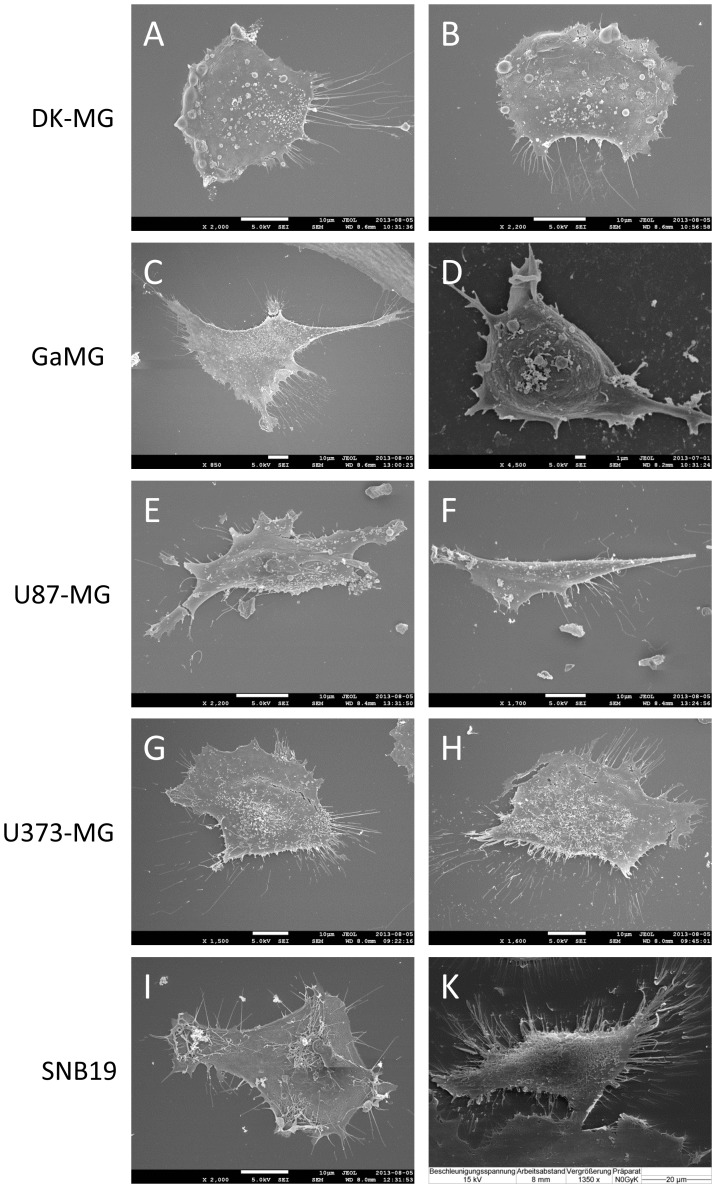
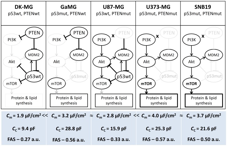
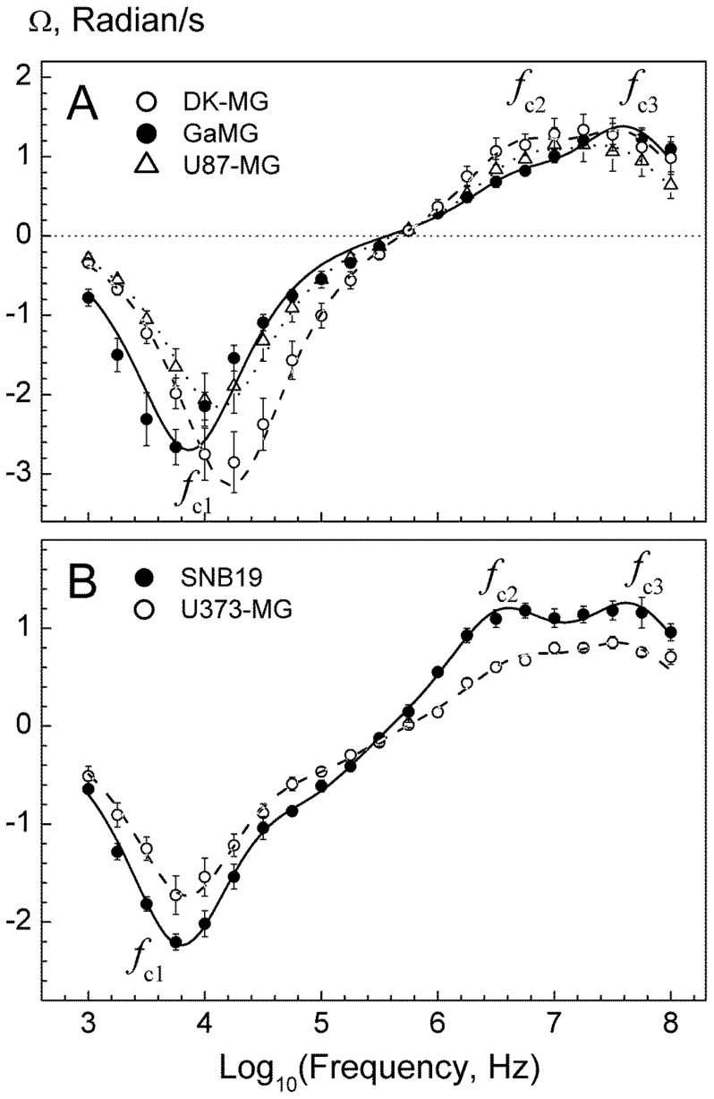
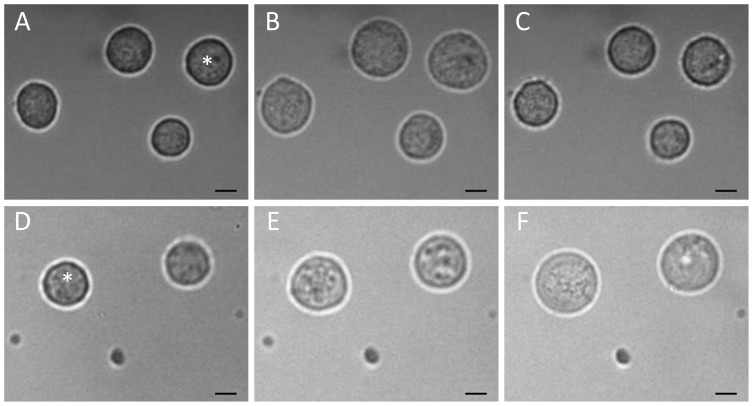
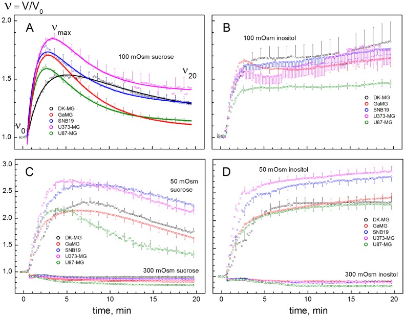
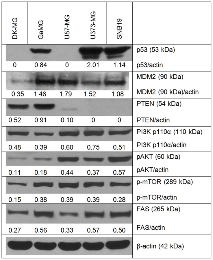
Glioblastoma multiforme (GBM) is characterized by rapid growth, invasion and resistance to chemo-/radiotherapy. The complex cell surface morphology with abundant membrane folds, microvilli, filopodia and other membrane extensions is believed to contribute to the highly invasive behavior and therapy resistance of GBM cells. The present study addresses the mechanisms leading to the excessive cell membrane area in five GBM lines differing in mutational status for PTEN and p53. In addition to scanning electron microscopy (SEM), the membrane area and folding were quantified by dielectric measurements of membrane capacitance using the single-cell electrorotation (ROT) technique. The osmotic stability and volume regulation of GBM cells were analyzed by video microscopy. The expression of PTEN, p53, mTOR and several other marker proteins involved in cell growth and membrane synthesis were examined by Western blotting. The combined SEM, ROT and osmotic data provided independent lines of evidence for a large variability in membrane area and folding among tested GBM lines. Thus, DK-MG cells (wild type p53 and wild type PTEN) exhibited the lowest degree of membrane folding, probed by the area-specific capacitance C m = 1.9 µF/cm(2). In contrast, cell lines carrying mutations in both p53 and PTEN (U373-MG and SNB19) showed the highest C m values of 3.7-4.0 µF/cm(2), which corroborate well with their heavily villated cell surface revealed by SEM. Since PTEN and p53 are well-known inhibitors of mTOR, the increased membrane area/folding in mutant GBM lines may be related to the enhanced protein and lipid synthesis due to a deregulation of the mTOR-dependent downstream signaling pathway. Given that membrane folds and extensions are implicated in tumor cell motility and metastasis, the dielectric approach presented here provides a rapid and simple tool for screening the biophysical cell properties in studies on targeting chemo- or radiotherapeutically the migration and invasion of GBM and other tumor types.
多形性胶质母细胞瘤(GBM)的特点是生长迅速、具有侵袭性且对化疗和放疗耐药。其复杂的细胞表面形态具有丰富的膜褶皱、微绒毛、丝状伪足和其他膜延伸结构,被认为有助于GBM细胞的高度侵袭行为和治疗抗性。本研究探讨了导致5种GBM细胞系细胞膜面积过大的机制,这些细胞系在PTEN和p53的突变状态上存在差异。除了扫描电子显微镜(SEM)外,还使用单细胞电旋转(ROT)技术通过测量膜电容的介电特性来量化膜面积和褶皱。通过视频显微镜分析GBM细胞的渗透稳定性和体积调节。通过蛋白质印迹法检测PTEN、p53、mTOR以及其他几种参与细胞生长和膜合成的标记蛋白的表达。SEM、ROT和渗透数据相结合,为测试的GBM细胞系之间膜面积和褶皱的巨大差异提供了独立的证据。因此,DK-MG细胞(野生型p53和野生型PTEN)表现出最低程度的膜褶皱,通过面积比电容(C_m) = 1.9 μF/cm²来衡量。相比之下,p53和PTEN均携带突变的细胞系(U373-MG和SNB19)显示出最高的(C_m)值,为3.7 - 4.0 μF/cm²,这与SEM显示的其高度绒毛状的细胞表面很好地吻合。由于PTEN和p53是众所周知的mTOR抑制剂,突变GBM细胞系中增加的膜面积/褶皱可能与mTOR依赖性下游信号通路失调导致的蛋白质和脂质合成增强有关。鉴于膜褶皱和延伸与肿瘤细胞的运动性和转移有关,本文介绍的介电方法为在针对GBM和其他肿瘤类型的迁移和侵袭进行化学或放射治疗的研究中筛选生物物理细胞特性提供了一种快速且简单的工具。